 Explore
 Validate
 Learn
Learn Flow cytometry
Antibody data
- Antibody Data
- Antigen structure
- References [3]
- Comments [0]
- Validations
- Flow cytometry [1]
- Other assay [2]
Submit
Validation data
Reference
Comment
Report error
- Product number
- 12-9863-42 - Provider product page

- Provider
- Invitrogen Antibodies
- Product name
- Phospho-NFkB p65 (Ser529) Monoclonal Antibody (B33B4WP), PE, eBioscience™
- Antibody type
- Monoclonal
- Antigen
- Other
- Description
- Description: The B33B4WP monoclonal antibody recognizes human NF kappa B (NFkB) p65 subunit when serine 529 is phosphorylated. NFkB, also known as nuclear factor kappa-light chain enhancer of activated B cells, is a ubiquitous transcription factor that regulates the transcription of many genes involved in cell proliferation, apoptosis, development, immunity and cancer. Functional NFkB is a homo- or hetero-dimer composed of 5 members of the NFkB family: p65 (RelA), c-Rel, RelB, p50 (NFkB1, p105 precursor protein), and p52 (NFkB2, p100 precursor protein). The activity of the complex is negatively regulated by binding to IkB inhibitors that sequester NFkB into the cytoplasm, inhibiting its transcriptional activity. NFkB-activating agents like tumor necrosis factor (TNF) alpha, interleukin-1 beta, lipopolysaccharide, camptothecin, and phorbol ester (PMA) induce the phosphorylation and degradation of IkB, leading to the translocation of NFkB to the nucleus where it binds to kB motifs and regulates gene expression. The activity of p65-containing NFkB complexes is positively regulated by phosphorylation of the p65 subunit at serine 529 and/or serine 536.
- Conjugate
- Yellow dye
- Antibody clone number
- B33B4WP
- Concentration
- 5 µL/Test
Submitted references S100A9 Derived From Myeloma Associated Myeloid Cells Promotes TNFSF13B/TNFRSF13B-Dependent Proliferation and Survival of Myeloma Cells.
Cas9-mediated excision of proximal DNaseI/H3K4me3 signatures confers robust silencing of microRNA and long non-coding RNA genes.
Human lactoferrin attenuates the proinflammatory response of neonatal monocyte-derived macrophages.
Meng L, Tang Q, Zhao J, Wang Z, Wei L, Wei Q, Yin L, Luo S, Song J
Frontiers in oncology 2021;11:691705
Frontiers in oncology 2021;11:691705
Cas9-mediated excision of proximal DNaseI/H3K4me3 signatures confers robust silencing of microRNA and long non-coding RNA genes.
Janga H, Aznaourova M, Boldt F, Damm K, Grünweller A, Schulte LN
PloS one 2018;13(2):e0193066
PloS one 2018;13(2):e0193066
Human lactoferrin attenuates the proinflammatory response of neonatal monocyte-derived macrophages.
Wisgrill L, Wessely I, Spittler A, Förster-Waldl E, Berger A, Sadeghi K
Clinical and experimental immunology 2018 Jun;192(3):315-324
Clinical and experimental immunology 2018 Jun;192(3):315-324
No comments: Submit comment
Supportive validation
- Submitted by
- Invitrogen Antibodies (provider)
- Main image

- Experimental details
- Intracellular staining of unstimulated (left) or 15-minute PMA-stimulated (right) normal human peripheral blood cells with Anti-Human CD57 FITC (Product # 11-0577-42) and Anti-Human phospho-NF kappa B p65 (S529) PE, using the Intracellular Fixation and Permeabilization Buffer Set (Product # 88-8824-00) and protocol. Cells in the lymphocyte gate were used for analysis.
- Conjugate
- Yellow dye

Supportive validation
- Submitted by
- Invitrogen Antibodies (provider)
- Main image

- Experimental details
- Figure 4 TNFSF13B/TNFRSF13B signal contributes to the canonical NF- k B signaling and the proliferation of MM cell. (A) Primary myeloma cells were derived from the patients with multiple myeloma. The expression of TNFRSF13B and TNFRSF17 on the myeloma cells. The data shown in the dot plot are the means +- SD of six samples from three independent experiments. (B) Myeloma cells were cultured with TNFSF13B either in the absence or presence of Bortezomib for 24 h. FACS plots indicate the proportion of KI67 + proliferative cells. The data shown in the dot plot are the means +- SD of six samples from three independent experiments. (C) The phosphorylation of p38 and (D) p65 has been demonstrated in FACS histogram after 30 min of treatment with TNFSF13B either in the presence or absence of Bortezomib. The data shown are the means +- SD of six samples from three independent experiments. ***P < 0.001.
- Conjugate
- Yellow dye

- Submitted by
- Invitrogen Antibodies (provider)
- Main image
- Experimental details
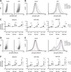
- Fig 4 Elevated NFkappaB p65 but not ERK1/2 activity on miR-146a and miR-155 knockout. A) Representative FACS scatter plots showing a right-shift of 30 min LPS-stimulated (1 mug / ml) compared to mock-treated monocytes stained with phospho-p65 antibody (PE-channel). B) Representative histogram plots showing an increased right-shift of miR-146a and miR-155 deficient compared to control or MALAT1 deficient monocytes after 30 min LPS-stimulation (1 mug / ml) and staining with a phospho-p65 antibody (PE-channel). C) Fold change in phospho-p65 signal in monocytes stimulated with LPS (1 mug / ml) for 15, 30 or 100 min compared to mock-treatment (ctrl) in wild-type (WT) or the indicated ncRNA knockout (KO) cells. All fold-changes are relative to the respective WT mock control. D-F) Same as A-C) but with phospho-ERK1/2 staining (APC-channel). Statistical significance was determined by a one-way ANOVA test with multiple comparisons (* p
- Conjugate
- Yellow dye
