Explore
 Validate
 Learn
Learn Western blot
 Immunocytochemistry
Antibody data
- Antibody Data
- Antigen structure
- References [3]
- Comments [0]
- Validations
- Western blot [2]
- Flow cytometry [1]
Submit
Validation data
Reference
Comment
Report error
- Product number
- PA5-16758 - Provider product page

- Provider
- Invitrogen Antibodies
- Product name
- Anti-NFkB p65 Polyclonal Antibody
- Antibody type
- Polyclonal
- Antigen
- Synthetic peptide
- Description
- PA5-16758 targets NFkappaB/p65 in IHC (P) and WB applications and shows reactivity with Human, Rabbit, and Rat samples. The PA5-16758 immunogen is a synthetic peptide corresponding to internal domain of human NFkappaB p65.
- Reactivity
- Human, Rat
- Host
- Rabbit
- Isotype
- IgG
- Vial size
- 500 µL
- Storage
- -20° C, Avoid Freeze/Thaw Cycles
Submitted references Pretreatment with mineralocorticoid receptor blocker reduces intestinal injury induced by ischemia and reperfusion: involvement of inhibition of inflammatory response, oxidative stress, nuclear factor κB, and inducible nitric oxide synthase.
NF-κB and COX-2 expression in nonmalignant endometrial lesions and cancer.
Mechanisms of the protective effects of curcumin against indomethacin-induced gastric ulcer in rats.
Ozacmak HS, Ozacmak VH, Barut F, Araslı M, Ucan BH
The Journal of surgical research 2014 Oct;191(2):350-61
The Journal of surgical research 2014 Oct;191(2):350-61
NF-κB and COX-2 expression in nonmalignant endometrial lesions and cancer.
Faloppa CC, Baiocchi G, Cunha IW, Fregnani JH, Osorio CA, Fukazawa EM, Kumagai LY, Badiglian-Filho L, Pinto GL, Soares FA
American journal of clinical pathology 2014 Feb;141(2):196-203
American journal of clinical pathology 2014 Feb;141(2):196-203
Mechanisms of the protective effects of curcumin against indomethacin-induced gastric ulcer in rats.
Morsy MA, El-Moselhy MA
Pharmacology 2013;91(5-6):267-74
Pharmacology 2013;91(5-6):267-74
No comments: Submit comment
Supportive validation
- Submitted by
- Invitrogen Antibodies (provider)
- Main image
- Experimental details
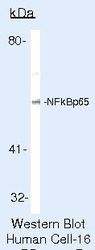
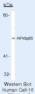
- Western blot of NFkappaB/p65 using NFkappaB/p65 Polyclonal Antibody (Product # PA5-16758) on LNCaP Cells.
- Submitted by
- Invitrogen Antibodies (provider)
- Main image
- Experimental details
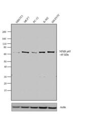
- Western blot analysis was performed using whole cell extracts (30 µg lysate) of NIH/3T3 (Lane 1), MCF7 (Lane 2), PC-12 (Lane 3), K-562 (Lane 4) and SH-SY5Y (Lane 5). The blots were probed with Anti-NFkappaB Rabbit polyclonal Antibody (Product # PA5-16758, 2-4 µg/mL) and detected by chemiluminescence using Goat anti-Rabbit IgG (H+L) Superclonal™ Secondary Antibody, HRP conjugate (Product # A27036, 0.4 µg/mL, 1:2500 dilution). A 65 kDa band corresponding to NFkappaB was observed across the cell lines tested. Known quantity of protein samples were electrophoresed using Novex® NuPAGE® 12 % Bis-Tris gel (Product # NP0342BOX), XCell SureLock™ Electrophoresis System (Product # EI0002) and Novex® Sharp Pre-Stained Protein Standard (Product # LC5800). Resolved proteins were then transferred onto a nitrocellulose membrane with iBlot® 2 Dry Blotting System (Product # IB21001). The membrane was probed with the relevant primary and secondary Antibody using iBind™ Flex Western Starter Kit (Product # SLF2000S). Chemiluminescent detection was performed using Pierce™ ECL Western Blotting Substrate (Product # 32106).

Supportive validation
- Submitted by
- Invitrogen Antibodies (provider)
- Main image
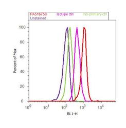
- Experimental details
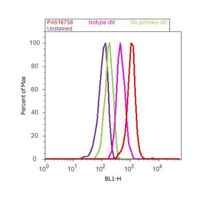
- Flow cytometry analysis of NFkappaB / p65 was performed using HeLa cells. Cells were fixed with 70% ethanol for 10 minutes, permeabilized with 0.25% Triton™ X-100 for 20 minutes, and blocked with 5% BSA for 30 minutes at room temperature. Cells were labeled with NFkappaB Rabbit Polyclonal Antibody (Product # PA5-16758, red histogram) or with rabbit isotype control (pink histogram) at 3-5 µg/million cells in 2.5% BSA. After incubation at room temperature for 2 hours, the cells were labeled with Alexa Fluor® 488 Goat Anti-Rabbit Secondary Antibody (Product # A11008) at a dilution of 1:400 for 30 minutes at room temperature. The representative 10, 000 cells were acquired and analyzed for each sample using an Attune® Acoustic Focusing Cytometer. The purple histogram represents unstained control cells and the green histogram represents no-primary-antibody control.