 Explore
 Validate
 Learn
Learn Western blot
 ELISA
ELISAAntibody data
- Antibody Data
- Antigen structure
- References [6]
- Comments [0]
- Validations
- Western blot [4]
- Immunohistochemistry [1]
Submit
Validation data
Reference
Comment
Report error
- Product number
- NBP1-77807 - Provider product page

- Provider
- Novus Biologicals
- Proper citation
- Novus Cat#NBP1-77807, RRID:AB_11021559
- Product name
- Rabbit Polyclonal RelA/NFkB p65 Antibody
- Antibody type
- Polyclonal
- Description
- Delipidation and Defibrination. This phospho specific polyclonal antibody is specific for phosphorylated pS276 human p65. Reactivity with non-phosphorylated p65 is minimal.
- Reactivity
- Human
- Host
- Rabbit
- Vial size
- 0.1 ml
- Storage
- Store at -20C. Avoid freeze-thaw cycles.
Submitted references Hunting for serine 276-phosphorylated p65.
CXCL5 promotes prostate cancer progression.
CXCL5 promotes prostate cancer progression.
Toll-like receptor 2 mediates apolipoprotein CIII-induced monocyte activation.
Piceatannol inhibits TNF-induced NF-kappaB activation and NF-kappaB-mediated gene expression through suppression of IkappaBalpha kinase and p65 phosphorylation.
Piceatannol inhibits TNF-induced NF-kappaB activation and NF-kappaB-mediated gene expression through suppression of IkappaBalpha kinase and p65 phosphorylation.
Spooren A, Kolmus K, Vermeulen L, Van Wesemael K, Haegeman G, Gerlo S
Journal of biomedicine & biotechnology 2010;2010:275892
Journal of biomedicine & biotechnology 2010;2010:275892
CXCL5 promotes prostate cancer progression.
Begley LA, Kasina S, Mehra R, Adsule S, Admon AJ, Lonigro RJ, Chinnaiyan AM, Macoska JA
Neoplasia (New York, N.Y.) 2008 Mar;10(3):244-54
Neoplasia (New York, N.Y.) 2008 Mar;10(3):244-54
CXCL5 promotes prostate cancer progression.
Begley LA, Kasina S, Mehra R, Adsule S, Admon AJ, Lonigro RJ, Chinnaiyan AM, Macoska JA
Neoplasia (New York, N.Y.) 2008 Mar;10(3):244-54
Neoplasia (New York, N.Y.) 2008 Mar;10(3):244-54
Toll-like receptor 2 mediates apolipoprotein CIII-induced monocyte activation.
Kawakami A, Osaka M, Aikawa M, Uematsu S, Akira S, Libby P, Shimokado K, Sacks FM, Yoshida M
Circulation research 2008 Dec 5;103(12):1402-9
Circulation research 2008 Dec 5;103(12):1402-9
Piceatannol inhibits TNF-induced NF-kappaB activation and NF-kappaB-mediated gene expression through suppression of IkappaBalpha kinase and p65 phosphorylation.
Ashikawa K, Majumdar S, Banerjee S, Bharti AC, Shishodia S, Aggarwal BB
Journal of immunology (Baltimore, Md. : 1950) 2002 Dec 1;169(11):6490-7
Journal of immunology (Baltimore, Md. : 1950) 2002 Dec 1;169(11):6490-7
No comments: Submit comment
Supportive validation
- Submitted by
- Novus Biologicals (provider)
- Main image

- Experimental details
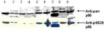
- Western Blot: RelA/NFkB p65 [p Ser276] Antibody [NBP1-77807] - pS529 shows phospho p65 staining in carcinoma cells.
- Submitted by
- Novus Biologicals (provider)
- Main image

- Experimental details
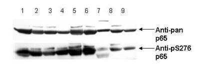
- Western Blot: RelA/NFkB p65 [p Ser276] Antibody [NBP1-77807] - pS276 shows phospho p65 staining in carcinoma cells. Western blot of total protein lysates from various human head and neck tumors shows phospho p65 staining in tumor cell lines using phospho specific polyclonal anti-human pS276 p65. Lanes 1-6 contain protein lysates from human squamal carcinoma cell lines. Lane 7 is a protein lysate from a primary culture of human keratinocytes and does not show significant levels of phosphorylated p65. Lane 8 contains protein lysate from ATCC SCC9 cells (also a head and neck squamal carcinoma). Lane 9 contains lysate from EGF-induced human derived A431 cells. Lane 10 (not shown) contains a molecular weight standard. Concurrent staining with anti-beta microtubulin (not shown) was used to confirm equal protein loading in all lanes. HRP conjugated Gt-anti-Rabbit IgG was used to develop the blot using a chemiluminescent detection method.
- Submitted by
- Novus Biologicals (provider)
- Main image
- Experimental details
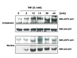
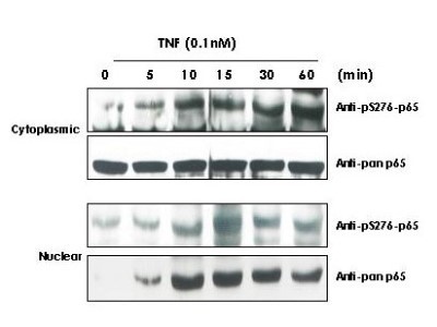
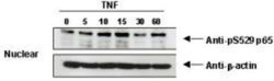
- Western Blot: RelA/NFkB p65 [p Ser276] Antibody [NBP1-77807] - TNF Induces phosphorylation of p65 in KBM-5 cells. Cytoplasmic and nuclear protein lysates prepared after 0, 5, 10, 15, 30 and 60 minutes of 0.1 nM TNF treatment of KBM-5 cells shows inducible phosphorylation using phospho specific polyclonal anti-human pS276 p65. Immunochemical's pan reactive anti p65 was used a control to show the presence of total p65 in both the cytoplasmic and nuclear extracts. Phosphorylation of p65 occurs after approximately 10 min of TNF exposure. Migration of phosphorylated p65 into the nucleus occurs within a similar time frame. HRP conjugated Gt-anti-Rabbit IgG was used to develop the blot using a chemi -luminescent detection method.
- Submitted by
- Novus Biologicals (provider)
- Main image
- Experimental details
- Western Blot: RelA/NFkB p65 [p Ser276] Antibody [NBP1-77807] - TNF Induces phosphorylation of p65 in KBM-5 cells.

Supportive validation
- Submitted by
- Novus Biologicals (provider)
- Main image
- Experimental details
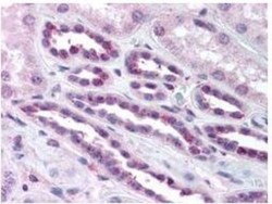
- Immunohistochemistry: RelA/NFkB p65 [p Ser276] Antibody [NBP1-77807] - Diluted to1:500 to detect p65 in human kidney tissue. Tissue was formalin fixed and paraffin embedded. No pre-treatment of sample was required. The image shows the localization of antibody as the precipitated red signal, with a hematoxylin purple nuclear counter stain.
