Explore
 Validate
 Learn
Learn Western blot
 ELISA
ELISAAntibody data
- Antibody Data
- Antigen structure
- References [7]
- Comments [0]
- Validations
- Western blot [3]
- Immunohistochemistry [1]
Submit
Validation data
Reference
Comment
Report error
- Product number
- NBP1-77808 - Provider product page

- Provider
- Novus Biologicals
- Proper citation
- Novus Cat#NBP1-77808, RRID:AB_11028293
- Product name
- Rabbit Polyclonal RelA/NFkB p65 Antibody
- Antibody type
- Polyclonal
- Description
- Delipidation and Defibrination. This phospho specific polyclonal antibody is specific for phosphorylated pS529 human p65. Reactivity with non-phosphorylated p65 is minimal.
- Reactivity
- Human
- Host
- Rabbit
- Vial size
- 0.1 ml
- Storage
- Store at -20C. Avoid freeze-thaw cycles.
Submitted references HIV-1 Tat-induced microgliosis and synaptic damage via interactions between peripheral and central myeloid cells.
Anti-inflammatory and anti-catabolic effects of TENDOACTIVE® on human tenocytes in vitro.
Role of NF-kappaB in flow-induced vascular remodeling.
The phosphatidylinositol 3-kinase/Akt pathway negatively regulates Nod2-mediated NF-kappaB pathway.
Suppression of NF-kappaB activation by curcumin leads to inhibition of expression of cyclo-oxygenase-2 and matrix metalloproteinase-9 in human articular chondrocytes: Implications for the treatment of osteoarthritis.
Piceatannol inhibits TNF-induced NF-kappaB activation and NF-kappaB-mediated gene expression through suppression of IkappaBalpha kinase and p65 phosphorylation.
Piceatannol inhibits TNF-induced NF-kappaB activation and NF-kappaB-mediated gene expression through suppression of IkappaBalpha kinase and p65 phosphorylation.
Lu SM, Tremblay MÈ, King IL, Qi J, Reynolds HM, Marker DF, Varrone JJ, Majewska AK, Dewhurst S, Gelbard HA
PloS one 2011;6(9):e23915
PloS one 2011;6(9):e23915
Anti-inflammatory and anti-catabolic effects of TENDOACTIVE® on human tenocytes in vitro.
Shakibaei M, Buhrmann C, Mobasheri A
Histology and histopathology 2011 Sep;26(9):1173-85
Histology and histopathology 2011 Sep;26(9):1173-85
Role of NF-kappaB in flow-induced vascular remodeling.
Castier Y, Ramkhelawon B, Riou S, Tedgui A, Lehoux S
Antioxidants & redox signaling 2009 Jul;11(7):1641-9
Antioxidants & redox signaling 2009 Jul;11(7):1641-9
The phosphatidylinositol 3-kinase/Akt pathway negatively regulates Nod2-mediated NF-kappaB pathway.
Zhao L, Lee JY, Hwang DH
Biochemical pharmacology 2008 Apr 1;75(7):1515-25
Biochemical pharmacology 2008 Apr 1;75(7):1515-25
Suppression of NF-kappaB activation by curcumin leads to inhibition of expression of cyclo-oxygenase-2 and matrix metalloproteinase-9 in human articular chondrocytes: Implications for the treatment of osteoarthritis.
Shakibaei M, John T, Schulze-Tanzil G, Lehmann I, Mobasheri A
Biochemical pharmacology 2007 May 1;73(9):1434-45
Biochemical pharmacology 2007 May 1;73(9):1434-45
Piceatannol inhibits TNF-induced NF-kappaB activation and NF-kappaB-mediated gene expression through suppression of IkappaBalpha kinase and p65 phosphorylation.
Ashikawa K, Majumdar S, Banerjee S, Bharti AC, Shishodia S, Aggarwal BB
Journal of immunology (Baltimore, Md. : 1950) 2002 Dec 1;169(11):6490-7
Journal of immunology (Baltimore, Md. : 1950) 2002 Dec 1;169(11):6490-7
No comments: Submit comment
Supportive validation
- Submitted by
- Novus Biologicals (provider)
- Main image
- Experimental details
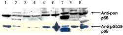
- Western Blot: RelA/NFkB p65 [p Ser529] Antibody [NBP1-77808] - pS529 shows phospho p65 staining in carcinoma cells. western blot of total protein lysates from various human head and neck tumors shows phospho p65 staining in tumor cell lines using phospho specific polyclonal anti-human pS529 p65. Lanes 1-6 contain protein lysates from human squamal carcinoma cell lines. Lane 7 is a protein lysate from a primary culture of human keratinocytes. Lane 8 contains protein lysate from ATCC SCC9 cells (also a head and neck squamal carcinoma). Lane 9 contains lysate from EGF-induced human derived A431 cells. Lane 10 (not shown) contains a molecular weight standard. Concurrent staining with anti-beta microtubulin (not shown) was used to confirm equal protein loading in all lanes. HRP conjugated Gt-anti-Rabbit IgG was used to develop the blot using a chemiluminescent detection method.

- Submitted by
- Novus Biologicals (provider)
- Main image
- Experimental details
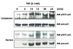
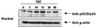
- Western Blot: RelA/NFkB p65 [p Ser529] Antibody [NBP1-77808] - TNF Induces phosphorylation of p65 in KBM-5 cells. Cytoplasmic and nuclear protein lysates prepared after 0, 5, 10, 15, 30 and 60 minutes of 0.1 nM TNF treatment of KBM-5 cells shows inducible phosphorylation using phospho specific polyclonal anti-human pS276 p65. Pan reactive anti p65 (NBP1-77808) was used a control to show the presence of total p65 in both the cytoplasmic and nuclear extracts. Phosphorylation of p65 occurs after approximately 10 min of TNF exposure. Migration of phosphorylated p65 into the nucleus occurs within a similar time frame. HRP conjugated Gt-anti-Rabbit IgG was used to develop the western blot using a chemi-luminescent detection method.

- Submitted by
- Novus Biologicals (provider)
- Main image
- Experimental details
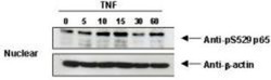
- Western Blot: RelA/NFkB p65 [p Ser529] Antibody [NBP1-77808] - 5 cells.
Supportive validation
- Submitted by
- Novus Biologicals (provider)
- Main image
- Experimental details
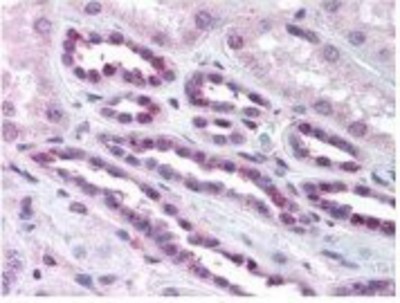
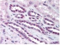
- Immunohistochemistry-Paraffin: RelA/NFkB p65 [p Ser529] Antibody [NBP1-77808] - pS276 shows phospho p65 staining in carcinoma cells.