Explore
 Validate
 Learn
Learn Western blot
Antibody data
- Antibody Data
- Antigen structure
- References [1]
- Comments [0]
- Validations
- Western blot [1]
- Immunohistochemistry [1]
- Other assay [1]
Submit
Validation data
Reference
Comment
Report error
- Product number
- PA5-23170 - Provider product page

- Provider
- Invitrogen Antibodies
- Product name
- NFkB p65 Polyclonal Antibody
- Antibody type
- Polyclonal
- Antigen
- Synthetic peptide
- Reactivity
- Human, Mouse, Bovine
- Host
- Rabbit
- Isotype
- IgG
- Vial size
- 100 µg
- Concentration
- 1.0 mg/mL
- Storage
- -20° C, Avoid Freeze/Thaw Cycles
Submitted references Baicalein ameliorates TNBS-induced colitis by suppressing TLR4/MyD88 signaling cascade and NLRP3 inflammasome activation in mice.
Luo X, Yu Z, Deng C, Zhang J, Ren G, Sun A, Mani S, Wang Z, Dou W
Scientific reports 2017 Nov 27;7(1):16374
Scientific reports 2017 Nov 27;7(1):16374
No comments: Submit comment
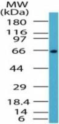
Supportive validation
- Submitted by
- Invitrogen Antibodies (provider)
- Main image
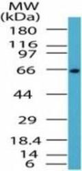
- Experimental details
- Western blot analysis of NFkB p65 in Jurkat lysate. Samples were incubated in NFkB p65 polyclonal antibody (Product # PA5-23170) using a dilution of 1 µg/mL followed by a goat anti-rabbit Ig HRP secondary antibody. PicoTect ECL substrate solution was used for this test.
Supportive validation
- Submitted by
- Invitrogen Antibodies (provider)
- Main image
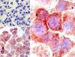
- Experimental details
- Immunohistochemical analysis of NFkB p65 in formalin-fixed, paraffin-embedded human HeLa cells (top left) and LPS-stimulated HeLa cells (bottom left, right). Samples were incubated in NFkB p65 polyclonal antibody (Product # PA5-23170) using a dilution of 5 µg/mL.

Supportive validation
- Submitted by
- Invitrogen Antibodies (provider)
- Main image

- Experimental details
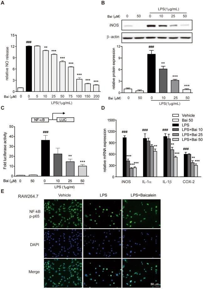
- Figure 4 Baicalein inhibited NF-kappaB pathway in vitro . Cells were treated with dose range of baicalein for 2 h prior to LPS (1 ug/ml) treatment for an additional 24 h. ( A ) The production of NO in RAW264.7 cells induced by LPS was measured as described in the Methods. ( B ) Protein level of RAW264.7 cells was determined with antibody against iNOS (1:1000) and beta-actin (1:2000 dilution) by immunoblotting. Quantification of the protein expression was performed by densitometric analysis of the blots. Expression was normalized to beta-actin. ( C ) NF-kappaB promoter-driven luciferase activity in RAW264.7 cells was determined using a luciferase assay system as described in the Methods. Results were expressed as fold values of control cells. ( D ) mRNA expression of iNOS, COX-2, IL-1alpha and IL-1beta in THP-1 cells was determined by qRT-PCR. Expression was normalized to beta-actin. ( E ) NF-kappaB p65 nuclear translocation in RAW264.7 cells was evaluated by immunofluorescence staining and images were captured by a fluorescence microscope. Scale bar corresponds to 50 mum and applies throughout. Data were expressed as mean +- SD of three independent experiments (n = 3). ### p < 0.001 vs. vehicle-treated group; **p < 0.01, ***P < 0.001 vs. LPS-treated group.