Explore
 Validate
 Learn
Learn Western blot
 Immunocytochemistry
 Immunoprecipitation
Antibody data
- Antibody Data
- Antigen structure
- References [3]
- Comments [0]
- Validations
- Immunocytochemistry [1]
- Other assay [2]
Submit
Validation data
Reference
Comment
Report error
- Product number
- PA5-17264 - Provider product page

- Provider
- Invitrogen Antibodies
- Product name
- Acetyl-NFkB p65 (Lys310) Polyclonal Antibody
- Antibody type
- Polyclonal
- Antigen
- Synthetic peptide
- Description
- It is not recommended to aliquot this antibody.
- Reactivity
- Human, Mouse
- Host
- Rabbit
- Isotype
- IgG
- Vial size
- 100 μL
- Concentration
- 62.1 μg/mL
- Storage
- -20°C
Submitted references Paeonol Protects Against Methotrexate-Induced Nephrotoxicity via Upregulation of P-gp Expression and Inhibition of TLR4/NF-κB Pathway.
Transforming Growth Factor β Inhibits MUC5AC Expression by Smad3/HDAC2 Complex Formation and NF-κB Deacetylation at K310 in NCI-H292 Cells.
Astaxanthin Ameliorates high-fat diet-induced cardiac damage and fibrosis by upregulating and activating SIRT1.
Morsy MA, El-Sheikh AAK, Abdel-Hafez SMN, Kandeel M, Abdel-Gaber SA
Frontiers in pharmacology 2022;13:774387
Frontiers in pharmacology 2022;13:774387
Transforming Growth Factor β Inhibits MUC5AC Expression by Smad3/HDAC2 Complex Formation and NF-κB Deacetylation at K310 in NCI-H292 Cells.
Lee SU, Kim MO, Kang MJ, Oh ES, Ro H, Lee RW, Song YN, Jung S, Lee JW, Lee SY, Bae T, Hong ST, Kim TD
Molecules and cells 2021 Jan 31;44(1):38-49
Molecules and cells 2021 Jan 31;44(1):38-49
Astaxanthin Ameliorates high-fat diet-induced cardiac damage and fibrosis by upregulating and activating SIRT1.
Shatoor AS, Al Humayed S
Saudi journal of biological sciences 2021 Dec;28(12):7012-7021
Saudi journal of biological sciences 2021 Dec;28(12):7012-7021
No comments: Submit comment
Supportive validation
- Submitted by
- Invitrogen Antibodies (provider)
- Main image

- Experimental details
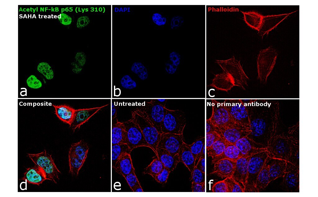
- Immunofluorescence analysis of Acetyl-NFkB p65 (Lys310) was performed using 70% confluent log phase HCT 116 cells treated with SAHA (2 micromolar) for 24 hours. The cells were fixed with 4% paraformaldehyde for 10 minutes, permeabilized with 0.1% Triton™ X-100 for 15 minutes, and blocked with 1% BSA for 1 hour at room temperature. The cells were labeled with Acetyl-NFkB p65 (Lys310) Rabbit Polyclonal Antibody (Product # PA5-17264) at 1:250 dilution in 0.1% BSA, incubated at 4 degree Celsius overnight and then labeled with Goat anti-Rabbit IgG (Heavy Chain) Superclonal™ Secondary Antibody, Alexa Fluor® 488 conjugate (Product # A27034) at a dilution of 1:2000 for 45 minutes at room temperature (Panel a: green). Nuclei (Panel b: blue) were stained with SlowFade® Gold Antifade Mountant with DAPI (Product # S36938). F-actin (Panel c: red) was stained with Rhodamine Phalloidin (Product # R415, 1:300). Panel d represents the merged image showing nuclear localization. Panel e shows untreated cells with no signal. Panel f represents control cells with no primary antibody to assess background. The images were captured at 60X magnification.
Supportive validation
- Submitted by
- Invitrogen Antibodies (provider)
- Main image
- Experimental details
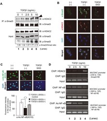
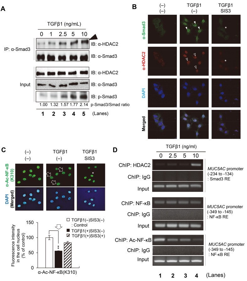
- Fig. 4 Smad3/HDAC2 complex inhibits NF-kappaB activation. (A) Co-immunoprecipitation (IP) using the Smad3 antibody. NCI-H292 cells were treated with the indicated concentrations of TGFbeta1 for 30 min and subsequently lysed for IP assays. The immunoprecipitants collected by the Smad3 antibody were immunoblotted (IB) with an antibody against HDAC2 or Smad3. The input panels represent 4% of the cell extracts used for IP, immunoblotted using antibodies against phosphorylated HDAC2, Smad3, and phosphorylated Smad3. TGFbeta-induced Smad3 phosphorylation increased in a concentration-dependent manner. Smad3 antibody was used as the loading control. The numbers at the bottom of each lane represent the relative band intensity normalized to the control (Smad3 without phosphorylation). (B) TGFbeta1-induced colocalization of SMAD3 and HDAC2 proteins in the cell nucleus (closed and open arrowheads, respectively). NCI-H292 cells were co-labeled using antibodies against Smad3 (green) and HDAC2 (red). The cell nucleus was stained with DAPI (blue). The merged confocal images were shown at the bottom. SIS3 (1 uM), a specific Smad3 inhibitor, was treated to suppress the nuclear colocalization of Smad3 or HDAC2 (asterisks). (C) The acetylated level at lysine 310 of NF-kappaB (Ac-NF-kappaB (K310)) was reduced by TGFbeta1 (arrows) and restored by an additional Smad3 inhibitor, SIS3 (asterisk). NCI-H292 cells were stained with anti-Ac-NF-kappaB (K310) antibody (green). Cell nuclei were stained with
- Submitted by
- Invitrogen Antibodies (provider)
- Main image

- Experimental details
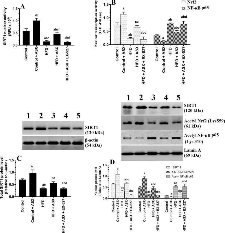
- Fig. 4 Nuclear activity of SIRT1 (A), the nuclear activity of Nrf2 and NF-kappaB p65 (B), total protein levels of SIRT1 (C), and nuclear protein levels of SIRT1, acetyl Nrf2, and acetyl NF-kappaB p65 in the left ventricles (LVs) of all groups of rats. Data were analyzed for n = 6 samples/group and were significantly different p < 0.05. a : vs. the control rats; b : vs. the control + ASX-treated rats; c : vs. HFD-fed rats, d : vs. HFD + ASX-treated rats. EX-527: a selective SIRT1 inhibitor.