Explore
 Validate
 Learn
Learn Western blot
Antibody data
- Antibody Data
- Antigen structure
- References [2]
- Comments [0]
- Validations
- Western blot [2]
- Immunocytochemistry [1]
- Flow cytometry [1]
Submit
Validation data
Reference
Comment
Report error
- Product number
- MAB5078 - Provider product page

- Provider
- R&D Systems
- Product name
- Human/Mouse/Rat RelA/NF kappa B p65 Antibody
- Antibody type
- Monoclonal
- Description
- Protein A or G purified from hybridoma culture supernatant. Detects human, mouse, and rat RelA/NF kappa B p65 in Western blots.
- Reactivity
- Human, Mouse, Rat
- Host
- Mouse
- Conjugate
- Unconjugated
- Antigen sequence
Q04206- Isotype
- IgG
- Antibody clone number
- 532301
- Vial size
- 100 ug
- Concentration
- LYOPH
- Storage
- Use a manual defrost freezer and avoid repeated freeze-thaw cycles. 12 months from date of receipt, -20 to -70 °C as supplied. 1 month, 2 to 8 °C under sterile conditions after reconstitution. 6 months, -20 to -70 °C under sterile conditions after reconstitution.
Submitted references Intermittent Hypoxia Activates Duration-Dependent Protective and Injurious Mechanisms in Mouse Lung Endothelial Cells.
The pro-inflammatory phenotype of the human non-classical monocyte subset is attributed to senescence.
Wohlrab P, Soto-Gonzales L, Benesch T, Winter MP, Lang IM, Markstaller K, Tretter V, Klein KU
Frontiers in physiology 2018;9:1754
Frontiers in physiology 2018;9:1754
The pro-inflammatory phenotype of the human non-classical monocyte subset is attributed to senescence.
Ong SM, Hadadi E, Dang TM, Yeap WH, Tan CT, Ng TP, Larbi A, Wong SC
Cell death & disease 2018 Feb 15;9(3):266
Cell death & disease 2018 Feb 15;9(3):266
No comments: Submit comment
Supportive validation
- Submitted by
- R&D Systems (provider)
- Main image

- Experimental details
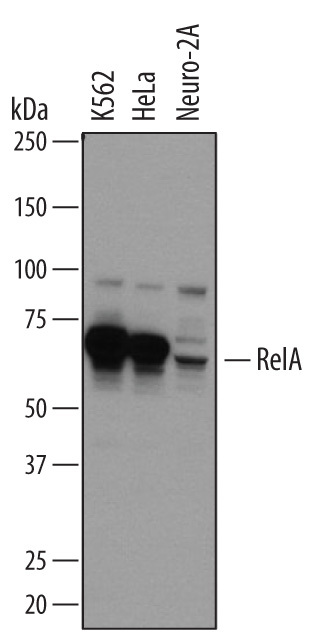
- Detection of Human and Mouse RelA/NF kappa B p65 by Western Blot. Western blot shows lysates of K562 human chronic myelogenous leukemia cell line, HeLa human cervical epithelial carcinoma cell line, and Neuro-2A mouse neuroblastoma cell line. PVDF membrane was probed with 2 µg/mL of Mouse Anti-Human/Mouse RelA/NF kappa B p65 Monoclonal Antibody (Catalog # MAB5078) followed by HRP-conjugated Anti-Mouse IgG Secondary Antibody (Catalog # HAF007). A specific band was detected for RelA/NF kappa B p65 at approximately 70 kDa (as indicated). This experiment was conducted under reducing conditions and using Immunoblot Buffer Group 1.
- Submitted by
- R&D Systems (provider)
- Main image

- Experimental details
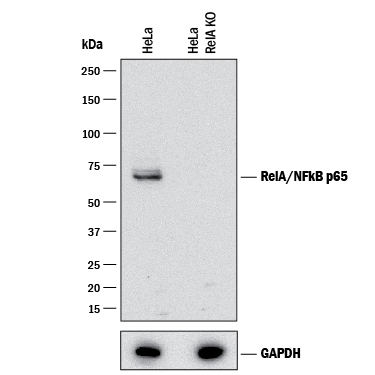
- Western Blot Shows Human RelA/NF kappa B p65 Specificity by Using Knockout Cell Line. Western blot shows lysates of HeLa human cervical epithelial carcinoma parental cell line and RelA/NF kappa B p65 knockout HeLa cell line (KO). PVDF membrane was probed with 2 µg/mL of Mouse Anti-Human/Mouse RelA/NF kappa B p65 Monoclonal Antibody (Catalog # MAB5078) followed by HRP-conjugated Anti-Mouse IgG Secondary Antibody (Catalog # HAF007). A specific band was detected for RelA/NF kappa B p65 at approximately 65 kDa (as indicated) in the parental HeLa cell line, but is not detectable in knockout HeLa cell line. GAPDH (Catalog # AF5718) is shown as a loading control. This experiment was conducted under reducing conditions and using Immunoblot Buffer Group 1.
Supportive validation
- Submitted by
- R&D Systems (provider)
- Main image

- Experimental details
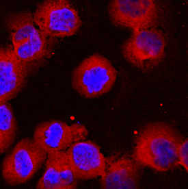
- RelA/NF kappa B p65 in K562 Human Cell Line. RelA/NF kappa B p65 was detected in immersion fixed K562 human chronic myelogenous leukemia cell line using Mouse Anti-Human/Mouse RelA/NF kappa B p65 Monoclonal Antibody (Catalog # MAB5078) at 10 µg/mL for 3 hours at room temperature. Cells were stained using the NorthernLights™ 557-conjugated Anti-Mouse IgG Secondary Antibody (red; Catalog # NL007) and counterstained with DAPI (blue). View our protocol for Fluorescent ICC Staining of Non-adherent Cells.
Supportive validation
- Submitted by
- R&D Systems (provider)
- Main image
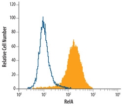
- Experimental details
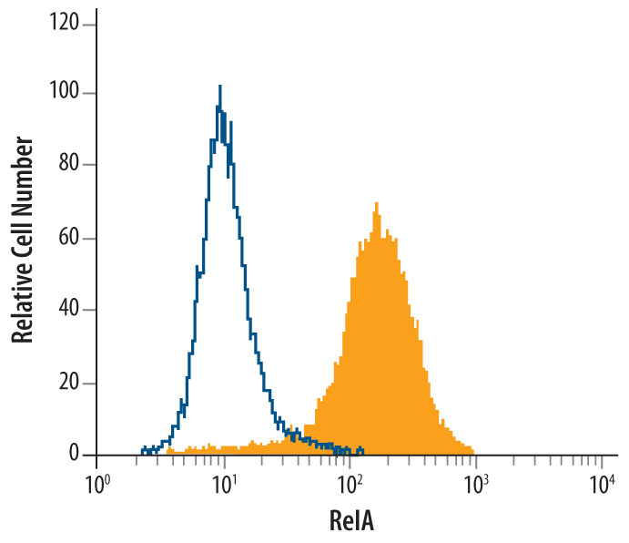
- Detection of RelA in HeLa Human Cell Line by Flow Cytometry. HeLa human cervical epithelial carcinoma cell line was stained with Mouse Anti-Human/Mouse RelA/NF kappa B p65 Monoclonal Antibody (Catalog # MAB5078, filled histogram) or isotype control antibody (Catalog # MAB0041, open histogram), followed by Phycoerythrin-conjugated Anti-Mouse IgG Secondary Antibody (Catalog # F0102B). To facilitate intracellular staining, cells were fixed with paraformadehyde and permeabilized with methanol.