Explore
 Validate
 Learn
Learn Western blot
 Immunocytochemistry
Antibody data
- Antibody Data
- Antigen structure
- References [9]
- Comments [0]
- Validations
- Western blot [10]
- Immunocytochemistry [1]
- Immunoprecipitation [2]
- Immunohistochemistry [3]
- Chromatin Immunoprecipitation [1]
Submit
Validation data
Reference
Comment
Report error
- Product number
- GTX101557 - Provider product page

- Provider
- GeneTex
- Proper citation
- GeneTex Cat#GTX101557, RRID:AB_1241080
- Product name
- mTOR antibody [C3], C-term
- Antibody type
- Polyclonal
- Reactivity
- Human, Mouse, Rat
- Host
- Rabbit
Submitted references Inflammatory interferon activates HIF-1α-mediated epithelial-to-mesenchymal transition via PI3K/AKT/mTOR pathway.
Activity-independent targeting of mTOR to lysosomes in primary osteoclasts.
The Inhibition of microRNA-128 on IGF-1-Activating mTOR Signaling Involves in Temozolomide-Induced Glioma Cell Apoptotic Death.
A microtubule inhibitor, ABT-751, induces autophagy and delays apoptosis in Huh-7 cells.
Proteome profiling of cadmium-induced apoptosis by antibody array analyses in human bronchial epithelial cells.
Prorenin receptor acts as a potential molecular target for pancreatic ductal adenocarcinoma diagnosis.
RNA-seq reveals aurora kinase-driven mTOR pathway activation in patients with sarcomatoid metastatic renal cell carcinoma.
Gab1 is essential for membrane translocation, activity and integrity of mTORCs after EGF stimulation in urothelial cell carcinoma.
Macrophage migration inhibitory factor induces vascular leakage via autophagy.
Yeh YH, Hsiao HF, Yeh YC, Chen TW, Li TK
Journal of experimental & clinical cancer research : CR 2018 Mar 27;37(1):70
Journal of experimental & clinical cancer research : CR 2018 Mar 27;37(1):70
Activity-independent targeting of mTOR to lysosomes in primary osteoclasts.
Wang A, Carraro-Lacroix LR, Owen C, Gao B, Corey PN, Tyrrell P, Brumell JH, Voronov I
Scientific reports 2017 Jun 7;7(1):3005
Scientific reports 2017 Jun 7;7(1):3005
The Inhibition of microRNA-128 on IGF-1-Activating mTOR Signaling Involves in Temozolomide-Induced Glioma Cell Apoptotic Death.
Chen PH, Cheng CH, Shih CM, Ho KH, Lin CW, Lee CC, Liu AJ, Chang CK, Chen KC
PloS one 2016;11(11):e0167096
PloS one 2016;11(11):e0167096
A microtubule inhibitor, ABT-751, induces autophagy and delays apoptosis in Huh-7 cells.
Wei RJ, Lin SS, Wu WR, Chen LR, Li CF, Chen HD, Chou CT, Chen YC, Liang SS, Chien ST, Shiue YL
Toxicology and applied pharmacology 2016 Nov 15;311:88-98
Toxicology and applied pharmacology 2016 Nov 15;311:88-98
Proteome profiling of cadmium-induced apoptosis by antibody array analyses in human bronchial epithelial cells.
Xu YM, Wu DD, Zheng W, Yu FY, Yang F, Yao Y, Zhou Y, Ching YP, Lau AT
Oncotarget 2016 Feb 2;7(5):6146-58
Oncotarget 2016 Feb 2;7(5):6146-58
Prorenin receptor acts as a potential molecular target for pancreatic ductal adenocarcinoma diagnosis.
Arundhathi A, Chuang WH, Chen JK, Wang SE, Shyr YM, Chen JY, Liao WN, Chen HW, Teng YM, Pai CC, Wang CH
Oncotarget 2016 Aug 23;7(34):55437-55448
Oncotarget 2016 Aug 23;7(34):55437-55448
RNA-seq reveals aurora kinase-driven mTOR pathway activation in patients with sarcomatoid metastatic renal cell carcinoma.
Pal SK, He M, Tong T, Wu H, Liu X, Lau C, Wang JH, Warden C, Wu X, Signoretti S, Choueiri TK, Karam JA, Jones JO
Molecular cancer research : MCR 2015 Jan;13(1):130-7
Molecular cancer research : MCR 2015 Jan;13(1):130-7
Gab1 is essential for membrane translocation, activity and integrity of mTORCs after EGF stimulation in urothelial cell carcinoma.
Chang CH, Chan PC, Li JR, Chen CJ, Shieh JJ, Fu YC, Chen HC, Wu MJ
Oncotarget 2015 Jan 30;6(3):1478-89
Oncotarget 2015 Jan 30;6(3):1478-89
Macrophage migration inhibitory factor induces vascular leakage via autophagy.
Chen HR, Chuang YC, Chao CH, Yeh TM
Biology open 2015 Jan 23;4(2):244-52
Biology open 2015 Jan 23;4(2):244-52
No comments: Submit comment
Enhanced validation
Supportive validation
- Submitted by
- GeneTex (provider)
- Enhanced method
- Genetic validation
- Main image
- Experimental details
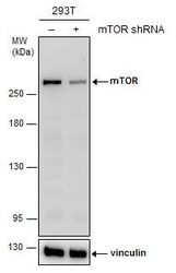
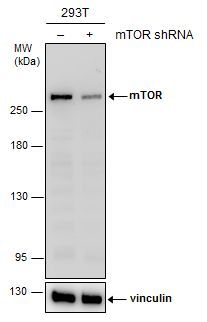
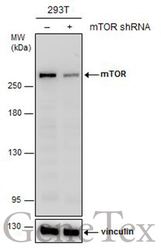
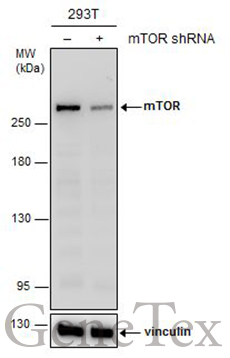
- Non-transfected (¡V) and transfected (+) 293T whole cell extracts (30 ?g) were separated by 5% SDS-PAGE, and the membrane was blotted with mTOR antibody [C3], C-term (GTX101557) diluted at 1:2000. The HRP-conjugated anti-rabbit IgG antibody (GTX213110-01) was used to detect the primary antibody.
Supportive validation
- Submitted by
- GeneTex (provider)
- Main image

- Experimental details
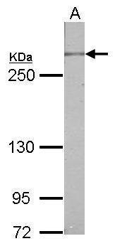
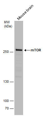
- Sample (50 ug of whole cell lysate) A: mouse brain 5% SDS PAGE GTX101557 diluted at 1:500
- Validation comment
- WB
- Submitted by
- GeneTex (provider)
- Main image
- Experimental details
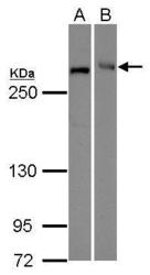
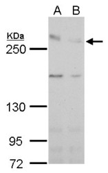
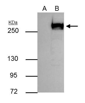
- mTOR antibody [C3], C-term detects FRAP1 protein by Western blot analysis.A. 30 ug 293T whole cell lysate/extractB. 30 ug Raji whole cell lysate/extract5 % SDS-PAGEmTOR antibody [C3], C-term (GTX101557) dilution: 1:1000
- Validation comment
- WB

- Submitted by
- GeneTex (provider)
- Main image
- Experimental details
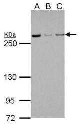
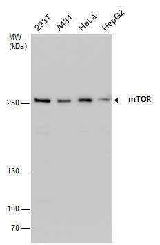
- mTOR antibody [C3], C-term detects FRAP1 protein by Western blot analysis.A. 30 µg 293T whole cell lysate/extract B. 30 µg A431 whole cell lysate/extract C. 30 µg HeLa whole cell lysate/extract5 % SDS-PAGEmTOR antibody [C3], C-term (GTX101557) dilution: 1:1000
- Validation comment
- WB
- Submitted by
- GeneTex (provider)
- Main image

- Experimental details
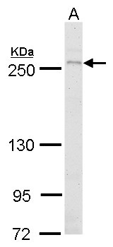
- mTOR antibody detects mTOR protein by western blot analysis.A. 50 ?g mouse brain lysate/extract5 % SDS-PAGEmTOR antibody (GTX101557) dilution: 1:500
- Validation comment
- WB
- Submitted by
- GeneTex (provider)
- Main image
- Experimental details
- mTOR antibody detects mTOR protein by western blot analysis.A. 30 ?g HeLa whole cell extract (untreated)B. 30 ?g HeLa whole cell extract (500 ?gM CoCl2 treatment for 24 hr)5 % SDS-PAGEmTOR antibody (GTX101557) dilution: 1:1000
- Validation comment
- WB

- Submitted by
- GeneTex (provider)
- Main image

- Experimental details
- mTOR antibody detects mTOR protein by western blot analysis.A. 30 ?g Raji whole cell lysate/extract5 % SDS-PAGEmTOR antibody (GTX101557) dilution: 1:1000
- Validation comment
- WB

- Submitted by
- GeneTex (provider)
- Main image
- Experimental details
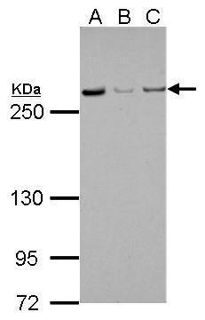
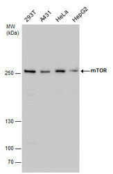
- mTOR antibody detects mTOR protein by western blot analysis. Various whole cell extracts (30 ?g) were separated by 5% SDS-PAGE, and the membrane was blotted with mTOR antibody (GTX101557) diluted by 1:1000. The HRP-conjugated anti-rabbit IgG antibody (GTX213110-01) was used to detect the primary antibody.
- Submitted by
- GeneTex (provider)
- Main image
- Experimental details
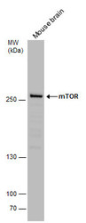
- mTOR antibody detects mTOR protein by western blot analysis. Mouse tissue extracts (50 ?g) was separated by 5% SDS-PAGE, and the membrane was blotted with mTOR antibody (GTX101557) diluted by 1:500. The HRP-conjugated anti-rabbit IgG antibody (GTX213110-01) was used to detect the primary antibody.
- Submitted by
- GeneTex (provider)
- Main image
- Experimental details
- Non-transfected (¡V) and transfected (+) 293T whole cell extracts (30 ?g) were separated by 5% SDS-PAGE, and the membrane was blotted with mTOR antibody [C3], C-term (GTX101557) diluted at 1:2000. The HRP-conjugated anti-rabbit IgG antibody (GTX213110-01) was used to detect the primary antibody.
Supportive validation
- Submitted by
- GeneTex (provider)
- Main image
- Experimental details
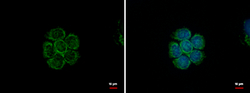
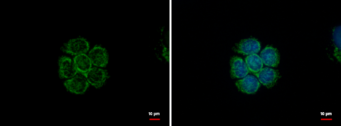
- mTOR antibody detects mTOR protein at cytoplasm and nucleus by immunofluorescent analysis. Sample: MCF-7 cells were fixed in 100% MeOH for 5 min.Green: mTOR protein stained by mTOR antibody (GTX101557) diluted at 1:500.Blue: Hoechst 33342 staining.
Supportive validation
- Submitted by
- GeneTex (provider)
- Main image

- Experimental details
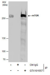
- mTOR antibody immunoprecipitates mTOR protein in IP experiments. IP Sample: 293T whole cell lysate/extract A : Control with 3 £gg of pre-immune rabbit IgG B : Immunoprecipitation of mTOR by 3 £gg of mTOR antibody (GTX101557) 5% SDS-PAGE The immunoprecipitated mTOR protein was detected by mTOR antibody (GTX101557) diluted at 1 : 1000. EasyBlot anti-rabbit IgG (HRP) (GTX221666-01) was used as a secondary reagent.
- Submitted by
- GeneTex (provider)
- Main image
- Experimental details
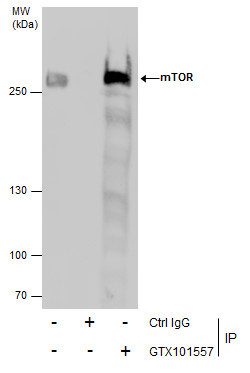
- Immunoprecipitation of mTOR protein from 293T whole cell extracts using 5 £gg of mTOR antibody [C3], C-term (GTX101557).Western blot analysis was performed using mTOR antibody [C3], C-term (GTX101557).EasyBlot anti-Rabbit IgG (GTX221666-01) was used as a secondary reagent.
Supportive validation
- Submitted by
- GeneTex (provider)
- Main image
- Experimental details
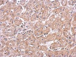
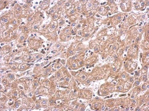
- mTOR antibody detects FRAP1 protein at cytosol on human hepatoma by immunohistochemical analysis. Sample: Paraffin-embedded hepatoma. mTOR antibody (GTX101557) dilution: 1:500.
- Submitted by
- GeneTex (provider)
- Main image
- Experimental details
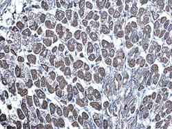
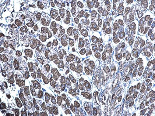
- mTOR antibody [C3], C-term detects mTOR protein at mitochondria on mouse stomach by immunohistochemical analysis. Sample: Paraffin-embedded mouse stomach. mTOR antibody [C3], C-term (GTX101557) diluted at 1:500.
- Submitted by
- GeneTex (provider)
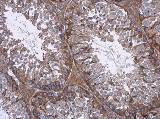
- Main image
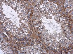
- Experimental details
- mTOR antibody [C3], C-term detects mTOR protein at cytoplasm in mouse testis by immunohistochemical analysis. Sample: Paraffin-embedded mouse testis. mTOR antibody [C3], C-term (GTX101557) diluted at 1:500.
Supportive validation
- Submitted by
- GeneTex (provider)
- Main image

- Experimental details
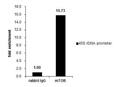
- Cross-linked ChIP was performed with 293T chromatin extract and 5 £gg of either control rabbit IgG or anti-mTOR antibody. The precipitated DNA was detected by PCR with primer set targeting to 45S rDNA promoter .