 Explore
 Validate
 Learn
LearnPA1-188
antibody from Invitrogen Antibodies
Targeting: MTOR
FLJ44809, FRAP, FRAP1, FRAP2, RAFT1, RAPT1
 Western blot
Antibody data
- Antibody Data
- Antigen structure
- References [0]
- Comments [0]
- Validations
- Western blot [2]
- Immunocytochemistry [2]
Submit
Validation data
Reference
Comment
Report error
- Product number
- PA1-188 - Provider product page

- Provider
- Invitrogen Antibodies
- Product name
- mTOR Polyclonal Antibody
- Antibody type
- Polyclonal
- Antigen
- Recombinant full-length protein
- Description
- Western blot analysis of PA1-188 detects mTOR at ~250 kDa in human and non-human primate cells, slightly below the theoretical MW of ~280kDa. Using a Tris-Acetate SDS gel for high molecular weight protein separation, and a nitrocellulose membrane for protein transfer are necessary for optimal WB results.
- Concentration
- 1 mg/mL
No comments: Submit comment
Supportive validation
- Submitted by
- Invitrogen Antibodies (provider)
- Main image

- Experimental details
- Western blot analysis of mTOR was performed by loading 50 µg of the indicated whole cell lysates per well, and 10 µL of HiMark™ Pre-stained Protein Standard onto a 3-8% NuPAGE® Tris-Acetate polyacrylamide gel. Proteins were transferred to a nitrocellulose membrane using the G2 Fast Blotter (Product # 62288), and blocked with StartingBlock T20 (TBS) Blocking Buffer (Product # 37543) for 1 hour at room temperature. mTOR was detected at ~250 kDa using an mTOR C-terminal polyclonal antibody (Product # PA1-188) at a dilution of 1:5000 in StartingBlock T20 (TBS) Blocking Buffer (Product # 37543) overnight at 4C on a rocking platform, followed by an HRP-conjugated goat anti-rabbit IgG secondary antibody (Product # 31460) at a dilution of 1:20,000 for 1 hour. Chemiluminescent detection was performed using SuperSignal West Dura (Product # 34075). Images were acquired on a Thermo Scientific myECL Imager (Product # 62236).

- Submitted by
- Invitrogen Antibodies (provider)
- Main image

- Experimental details
- CRISPR-Cas9 mediated genome editing ofmTOR (as confirmed by next generation sequencing) was achieved by using LentiArray™ Lentiviral sgRNA (Product # A32042, AssayIDCRISPR814789_LV) and LentiArray Cas9 Lentivirus (Product # A32064). Fig (a) Western blot analysis of mTOR was performed by loading 30 µg of HeLa Wild Type (Lane 1), HeLa Cas9 (Lane 2) and HeLa Cas9 cells transduced with mTOR Lentiviral sgRNA (Lane 3) whole cell extracts. The samples were electrophoresed using NuPAGE™ 3 to 8%, Tris-Acetate, 1.0 mm, Mini Protein Gel (Product # EC6695BOX). Resolved proteins were then transferred onto a nitrocellulose membrane (Product # IB23001) by iBlot® 2 Dry Blotting System (Product # IB21001). The blot was probed with Anti-mTOR Polyclonal Antibody (Product # PA1-188) using 1:5000 dilution and Goat anti-Rabbit IgG (H+L) Superclonal™ Recombinant Secondary Antibody, HRP (Product # A27036 1:20000 dilution).Chemiluminescent detection was performed using SuperSignal™ West Atto Ultimate Sensitivity Substrate (Product # A38556). A reduced signal in sgRNA transduced cells using the LentiArray™ CRISPR product line confirms that antibody is specific tomTOR (Fig (b)).

Supportive validation
- Submitted by
- Invitrogen Antibodies (provider)
- Main image
- Experimental details
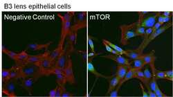
- Immunofluorescent analysis of mTOR (green) in B3 cells. The cells were fixed with formaldehyde for 15 minutes, permeabilized with 0.1% Triton X-100 in TBS for 10 minutes, and blocked with 1% Blocker BSA in PBS (Product # 37525) for 15 minutes, all at room temperature. Cells were stained with an mTOR C-terminal polyclonal antibody (Product # PA1-188) at a dilution of 1:50 in 1% Blocker BSA in PBS (Product # 37525) for 1 hour at room temperature, and then incubated with a DyLight 488-conjugated goat anti-rabbit IgG secondary antibody (Product # 35552) at a dilution of 1:250 for 30 minutes at room temperature. F-Actin (red) was stained with DyLight-554 Phalloidin (Product # 21834) and nuclei (blue) were stained with Hoechst 33342 dye (Product # 62249). Images were taken on a Thermo Scientific ToxInsight Instrument at 20X magnification.

- Submitted by
- Invitrogen Antibodies (provider)
- Main image
- Experimental details
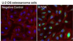
- Immunofluorescent analysis of mTOR (green) in U-2 OS cells. The cells were fixed with formaldehyde for 15 minutes, permeabilized with 0.1% Triton X-100 in TBS for 10 minutes, and blocked with 1% Blocker BSA in PBS (Product # 37525) for 15 minutes, all at room temperature. Cells were stained with an mTOR C-terminal polyclonal antibody (Product # PA1-188) at a dilution of 1:50 in 1% Blocker BSA in PBS (Product # 37525) for 1 hour at room temperature, and then incubated with a DyLight 488-conjugated goat anti-rabbit IgG secondary antibody (Product # 35552) at a dilution of 1:250 for 30 minutes at room temperature. F-Actin (red) was stained with DyLight-554 Phalloidin (Product # 21834) and nuclei (blue) were stained with Hoechst 33342 dye (Product # 62249). Images were taken on a Thermo Scientific ToxInsight Instrument at 20X magnification.
