Explore
 Validate
 Learn
LearnPA1-518
antibody from Invitrogen Antibodies
Targeting: MTOR
FLJ44809, FRAP, FRAP1, FRAP2, RAFT1, RAPT1
 Western blot
 Immunocytochemistry
Antibody data
- Antibody Data
- Antigen structure
- References [2]
- Comments [0]
- Validations
- Immunocytochemistry [2]
- Immunoprecipitation [1]
- Other assay [2]
Submit
Validation data
Reference
Comment
Report error
- Product number
- PA1-518 - Provider product page

- Provider
- Invitrogen Antibodies
- Product name
- mTOR Polyclonal Antibody
- Antibody type
- Polyclonal
- Antigen
- Other
- Description
- 3-8% Tris-Acetate Gel is recommended for Western blot. PA1-518 detects a predominant band at ~290kD corresponding to mTOR. Other lower MW nonspecific bands of unknown identity were also detected at ~65kD and ~35kD in some cell lysates.
- Reactivity
- Human, Mouse, Rat
- Host
- Rabbit
- Isotype
- IgG
- Vial size
- 100 μg
- Concentration
- 1 mg/mL
- Storage
- -20°C
Submitted references Effect and molecular mechanism of mTOR inhibitor rapamycin on temozolomide-induced autophagic death of U251 glioma cells.
Bilirubin activates transcription of HIF-1α in human proximal tubular cells cultured in the physiologic oxygen content.
Li B, Zhou C, Yi L, Xu L, Xu M
Oncology letters 2018 Feb;15(2):2477-2484
Oncology letters 2018 Feb;15(2):2477-2484
Bilirubin activates transcription of HIF-1α in human proximal tubular cells cultured in the physiologic oxygen content.
Kim SG, Ahn SY, Lee ES, Kim S, Na KY, Chae DW, Chin HJ
Journal of Korean medical science 2014 Sep;29 Suppl 2(Suppl 2):S146-54
Journal of Korean medical science 2014 Sep;29 Suppl 2(Suppl 2):S146-54
No comments: Submit comment
Supportive validation
- Submitted by
- Invitrogen Antibodies (provider)
- Main image
- Experimental details
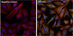
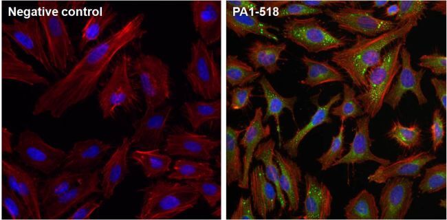
- Immunofluorescent analysis of mTOR (green) in HeLa cells. The cells were fixed with 4% paraformaldehyde, permeabilized with 0.1% Triton X-100 in PBS, and blocked with 1% Blocker BSA (Product # 37525), each for 15 minutes at room temperature. Cells were stained with an mTOR polyclonal antibody (Product # PA1-518) at a concentration of 10 µg/mL in 1% Blocker BSA in PBS (right panel), or incubated in blocking buffer alone as a negative control (left panel) overnight at 4C, and then incubated with a Dylight 488-conjugated goat anti-rabbit IgG secondary antibody (Product # 35552) at a dilution of 1:1000 for 1 hour at room temperature. F-Actin (red) was stained with a DyLight 554-conjugated phalloidin control (Product # 21834) and nuclei (blue) were stained with DAPI (Product # 46190).
- Submitted by
- Invitrogen Antibodies (provider)
- Main image
- Experimental details
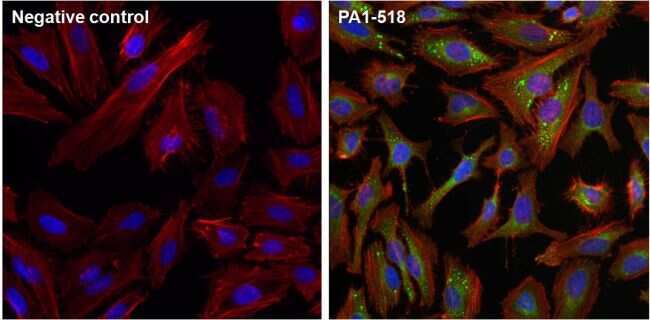
- Immunofluorescent analysis of mTOR (green) in HeLa cells. The cells were fixed with 4% paraformaldehyde, permeabilized with 0.1% Triton X-100 in PBS, and blocked with 1% Blocker BSA (Product # 37525), each for 15 minutes at room temperature. Cells were stained with an mTOR polyclonal antibody (Product # PA1-518) at a concentration of 10 µg/mL in 1% Blocker BSA in PBS (right panel), or incubated in blocking buffer alone as a negative control (left panel) overnight at 4C, and then incubated with a Dylight 488-conjugated goat anti-rabbit IgG secondary antibody (Product # 35552) at a dilution of 1:1000 for 1 hour at room temperature. F-Actin (red) was stained with a DyLight 554-conjugated phalloidin control (Product # 21834) and nuclei (blue) were stained with DAPI (Product # 46190).
Supportive validation
- Submitted by
- Invitrogen Antibodies (provider)
- Main image

- Experimental details
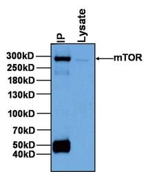
- Immunoprecipitation of mTOR was performed on HeLa cells. Antigen-antibody complexes were formed by incubating 500 µg of HeLa whole cell lysate with 2 µg of mTOR polyclonal antibody (Product # PA1-518) overnight on a rocking platform at 4C. The immune complexes were captured on 50 µL of Protein A/G Agarose (Product # 20421), washed extensively, and eluted with Lane Marker Reducing Sample Buffer (Product # 39000). HeLa cell lysate (10 µg) was loaded as a positive control (right lane). Samples were resolved on a NuPAGE® 3-8% Tris-Acetate Gel, transferred to a PVDF membrane, and blocked with StartingBlock T20 (TBS) Blocking Buffer (Product # 37543) for 1 hour at room temperature. The membrane was probed with an mTOR polyclonal antibody (Product # PA1-518) at a concentration of 6.5 µg/mL in StartingBlock T20 (TBS) Blocking Buffer (Product # 37543) overnight at 4C on a rocking platform, washed in TBST, and probed with an HRP-conjugated goat anti-rabbit IgG secondary antibody (Product # 31460) at a dilution of 1:40,000 for 1 hour. Chemiluminescent detection was performed using SuperSignal West Pico (Product # 34080).

Supportive validation
- Submitted by
- Invitrogen Antibodies (provider)
- Main image
- Experimental details
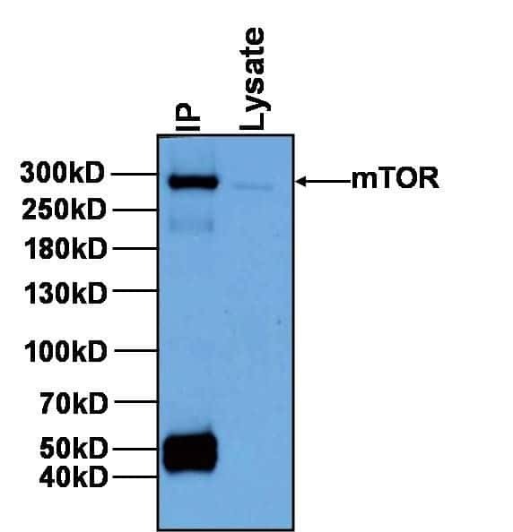
- Immunoprecipitation of mTOR was performed on HeLa cells. Antigen-antibody complexes were formed by incubating 500 µg of HeLa whole cell lysate with 2 µg of mTOR polyclonal antibody (Product # PA1-518) overnight on a rocking platform at 4C. The immune complexes were captured on 50 µL of Protein A/G Agarose (Product # 20421), washed extensively, and eluted with Lane Marker Reducing Sample Buffer (Product # 39000). HeLa cell lysate (10 µg) was loaded as a positive control (right lane). Samples were resolved on a NuPAGE? 3-8% Tris-Acetate Gel, transferred to a PVDF membrane, and blocked with StartingBlock T20 (TBS) Blocking Buffer (Product # 37543) for 1 hour at room temperature. The membrane was probed with an mTOR polyclonal antibody (Product # PA1-518) at a concentration of 6.5 µg/mL in StartingBlock T20 (TBS) Blocking Buffer (Product # 37543) overnight at 4C on a rocking platform, washed in TBST, and probed with an HRP-conjugated goat anti-rabbit IgG secondary antibody (Product # 31460) at a dilution of 1:40,000 for 1 hour. Chemiluminescent detection was performed using SuperSignal West Pico (Product # 34080).
- Submitted by
- Invitrogen Antibodies (provider)
- Main image
- Experimental details
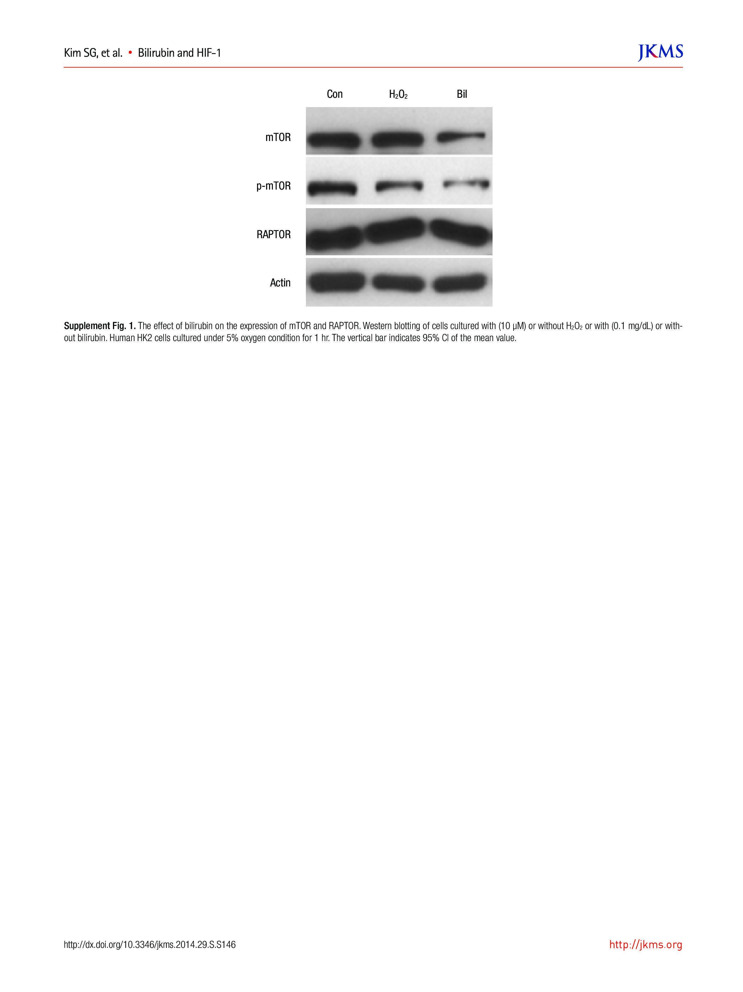
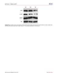
- Supplement Fig. 1 The effect of bilirubin on the expression of mTOR and RAPTOR. Western blotting of cells cultured with (10 uM) or without H 2 O 2 or with (0.1 mg/dL) or without bilirubin. Human HK2 cells cultured under 5% oxygen condition for 1 hr. The vertical bar indicates 95% CI of the mean value.