PA5-17780
antibody from Invitrogen Antibodies
Targeting: MTOR
FLJ44809, FRAP, FRAP1, FRAP2, RAFT1, RAPT1
Antibody data
- Antibody Data
- Antigen structure
- References [2]
- Comments [0]
- Validations
- Western blot [2]
- Other assay [1]
Submit
Validation data
Reference
Comment
Report error
- Product number
- PA5-17780 - Provider product page
- Provider
- Invitrogen Antibodies
- Product name
- mTOR Polyclonal Antibody
- Antibody type
- Polyclonal
- Antigen
- Synthetic peptide
- Description
- It is not recommended to aliquot this antibody.
- Reactivity
- Human, Mouse, Rat
- Host
- Rabbit
- Isotype
- IgG
- Vial size
- 100 µL
- Concentration
- 33 µg/mL
- Storage
- -20°C
Submitted references Space radiation exposure persistently increased leptin and IGF1 in serum and activated leptin-IGF1 signaling axis in mouse intestine.
Radiation persistently promoted oxidative stress, activated mTOR via PI3K/Akt, and downregulated autophagy pathway in mouse intestine.
Suman S, Kumar S, Fornace AJ, Datta K
Scientific reports 2016 Aug 25;6:31853
Radiation persistently promoted oxidative stress, activated mTOR via PI3K/Akt, and downregulated autophagy pathway in mouse intestine.
Datta K, Suman S, Fornace AJ Jr
The international journal of biochemistry & cell biology 2014 Dec;57:167-76
No comments: Submit comment
Supportive validation
- Submitted by
- Invitrogen Antibodies (provider)
- Main image
- Experimental details
- Western blot analysis of FRAP1 was performed with 10 µg of A549 cells transfected with Transfection Reagent alone (Lane 1), 100nM Non-Targeting control siRNA (Lane 2), or 100nM siRNA against FRAP1 (Lane 3). Proteins were resolved using a NuPAGE® Novex 4-12% Bis-Tris Gel (Product # NP0322BOX), XCell SureLock™ Electrophoresis System (Product # EI0002), and a protein size ladder. Proteins were wet transferred to a Pierce Nitrocellulose Membrane (Product # 88025) OR Pierce PVDF Membrane (Product # 88518) and blocked with Pierce Starting Block T20 (PBS) Blocking Buffer (Product # 37539) for 1 hour at room temperature. FRAP1 was detected at ~ 289 kDa using FRAP1 Rabbit polyclonal antibody (Product # PA5-17780) diluted in Pierce Starting Block T20 (PBS) Blocking Buffer 4°C overnight on a rocking platform. Pierce Goat Anti-Rabbit (Product # 31461) HRP-Conjugated Antibodies at a 1:2500 dilution were used and chemiluminescent detection was performed using Pierce Supersignal West Dura Maximum Sensitivity Substrate (Product # 37071). Relative density of the bands normalized to GAPDH (36 kDa). FRAP1 Antibody (Product # PA5-17780) confirms silencing of FRAP1 expression.
Supportive validation
- Submitted by
- Invitrogen Antibodies (provider)
- Main image
- Experimental details
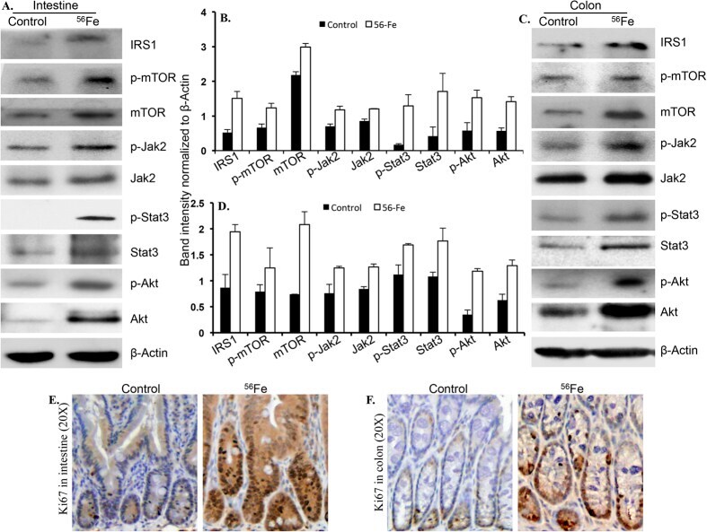
- Figure 4 Immunoblot analysis of IRS1, p-mTOR, mTOR, p-Jak2, Jak2, p-Stat3, Stat3, p-Akt, Akt showed activation of IGF1 and leptin signaling in intestine ( A,B ) and colon ( C,D ) 12 months after radiation exposure. Immunostaining of Ki-67 in small intestine sections from irradiated mice showed greater staining relative to control mice ( A,B ). Colon sections from irradiated mice also showed increased Ki67 staining relative to controls ( C,D ). Error bars represent mean +- SEM and p < 0.05 was considered significant, compared to sham irradiated control.