Explore
 Validate
 Learn
Learn Western blot
 Immunocytochemistry
Antibody data
- Antibody Data
- Antigen structure
- References [3]
- Comments [0]
- Validations
- Western blot [6]
- Immunocytochemistry [1]
- Immunoprecipitation [1]
- Immunohistochemistry [1]
- Chromatin Immunoprecipitation [1]
Submit
Validation data
Reference
Comment
Report error
- Product number
- GTX113303 - Provider product page

- Provider
- GeneTex
- Proper citation
- GeneTex Cat#GTX113303, RRID:AB_10721050
- Product name
- HDAC3 antibody
- Antibody type
- Polyclonal
- Reactivity
- Human, Mouse, Rat
- Host
- Rabbit
Submitted references An HDAC3-PROX1 corepressor module acts on HNF4α to control hepatic triglycerides.
Histone deacetylase 3 prepares brown adipose tissue for acute thermogenic challenge.
In cortical neurons HDAC3 activity suppresses RD4-dependent SMRT export.
Armour SM, Remsberg JR, Damle M, Sidoli S, Ho WY, Li Z, Garcia BA, Lazar MA
Nature communications 2017 Sep 15;8(1):549
Nature communications 2017 Sep 15;8(1):549
Histone deacetylase 3 prepares brown adipose tissue for acute thermogenic challenge.
Emmett MJ, Lim HW, Jager J, Richter HJ, Adlanmerini M, Peed LC, Briggs ER, Steger DJ, Ma T, Sims CA, Baur JA, Pei L, Won KJ, Seale P, Gerhart-Hines Z, Lazar MA
Nature 2017 Jun 22;546(7659):544-548
Nature 2017 Jun 22;546(7659):544-548
In cortical neurons HDAC3 activity suppresses RD4-dependent SMRT export.
Soriano FX, Hardingham GE
PloS one 2011;6(6):e21056
PloS one 2011;6(6):e21056
No comments: Submit comment
Enhanced validation
Supportive validation
- Submitted by
- GeneTex (provider)
- Enhanced method
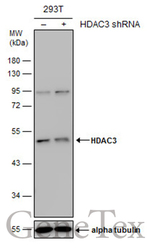
- Genetic validation
- Main image
- Experimental details
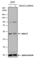
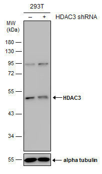
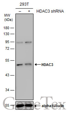
- Non-transfected (¡V) and transfected (+) 293T whole cell extracts (30 ?g) were separated by 10% SDS-PAGE, and the membrane was blotted with HDAC3 antibody (GTX113303) diluted at 1:500. The HRP-conjugated anti-rabbit IgG antibody (GTX213110-01) was used to detect the primary antibody.
Supportive validation
- Submitted by
- GeneTex (provider)
- Main image
- Experimental details
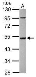
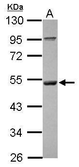
- Sample (30 ug of whole cell lysate) A: A431 10% SDS PAGE GTX113303 diluted at 1:1000
- Validation comment
- WB
- Submitted by
- GeneTex (provider)
- Main image
- Experimental details
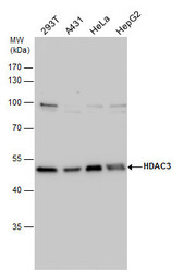
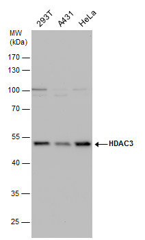
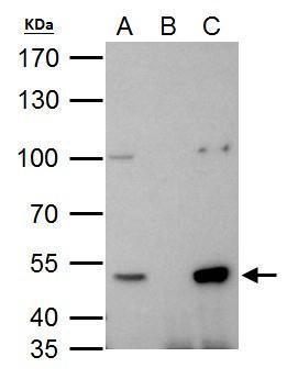
- HDAC3 antibody detects HDAC3 protein by western blot analysis. Various whole cell extracts (30 ?g) were separated by 10% SDS-PAGE, and the membrane was blotted with HDAC3 antibody (GTX113303) diluted by 1:1000. The HRP-conjugated anti-rabbit IgG antibody (GTX213110-01) was used to detect the primary antibody.
- Submitted by
- GeneTex (provider)
- Main image
- Experimental details
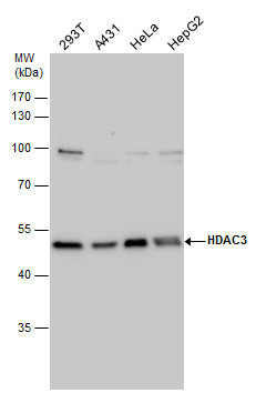
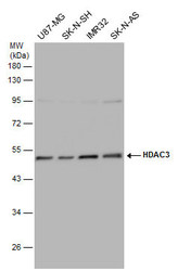
- HDAC3 antibody detects HDAC3 protein by western blot analysis. Various whole cell extracts (30 ?g) were separated by 10% SDS-PAGE, and the membrane was blotted with HDAC3 antibody (GTX113303) diluted by 1:1000.
- Validation comment
- WB
- Submitted by
- GeneTex (provider)
- Main image
- Experimental details
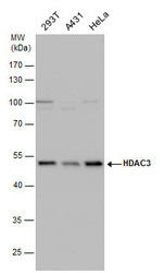
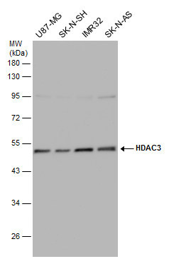
- Various whole cell extracts (30 ?g) were separated by 10% SDS-PAGE, and the membrane was blotted with HDAC3 antibody (GTX113303) diluted at 1:500. The HRP-conjugated anti-rabbit IgG antibody (GTX213110-01) was used to detect the primary antibody.
- Submitted by
- GeneTex (provider)
- Main image
- Experimental details
- Non-transfected (¡V) and transfected (+) 293T whole cell extracts (30 ?g) were separated by 10% SDS-PAGE, and the membrane was blotted with HDAC3 antibody (GTX113303) diluted at 1:500. The HRP-conjugated anti-rabbit IgG antibody (GTX213110-01) was used to detect the primary antibody.
Supportive validation
- Submitted by
- GeneTex (provider)
- Main image
- Experimental details
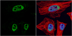
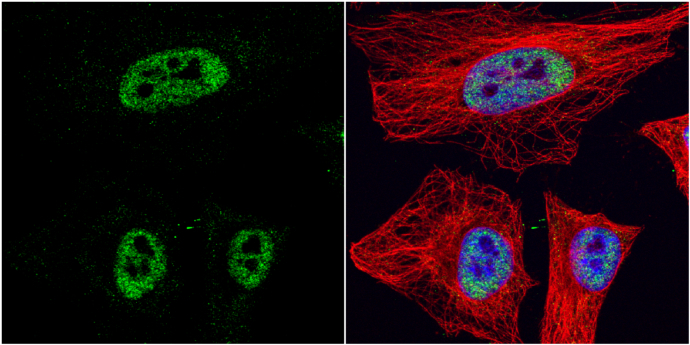
- HDAC3 antibody detects HDAC3 protein at nucleus by immunofluorescent analysis.Sample: HeLa cells were fixed in 4% paraformaldehyde at RT for 15 min.Green: HDAC3 protein stained by HDAC3 antibody (GTX113303) diluted at 1:1000.Red: alpha Tubulin, a cytoskeleton marker, stained by alpha Tubulin antibody [GT114] (GTX628802) diluted at 1:1000.Blue: Hoechst 33342 staining.
Supportive validation
- Submitted by
- GeneTex (provider)
- Main image
- Experimental details
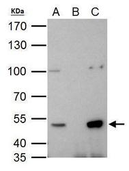
- HDAC3 antibody immunoprecipitates HDAC3 protein in IP experiments. IP Sample: 293T whole cell lysate/extract A. 40 £gg 293T whole cell lysate/extract B. Control with 2 £gg of preimmune rabbit IgG C. Immunoprecipitation of HDAC3 protein by 2 £gg of HDAC3 antibody (GTX113303) 7.5% SDS-PAGE The immunoprecipitated HDAC3 protein was detected by HDAC3 antibody (GTX113303) diluted at 1:1000. EasyBlot anti-rabbit IgG (GTX221666-01) was used as a secondary reagent.
Supportive validation
- Submitted by
- GeneTex (provider)
- Main image
- Experimental details
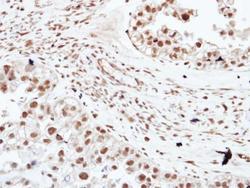
- Immunohistochemical analysis of paraffin-embedded human ovarian cancer, using HDAC3(GTX113303) antibody at 1:250 dilution.

Supportive validation
- Submitted by
- GeneTex (provider)
- Main image

- Experimental details
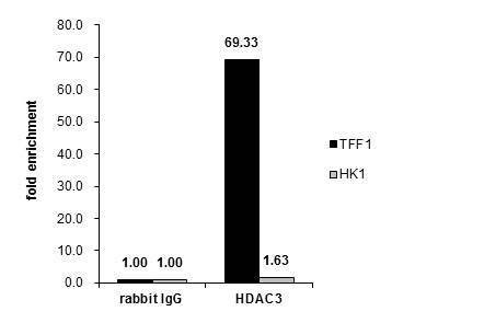
- Cross-linked ChIP was performed with MCF-7 chromatin extract and 5 £gg of either control rabbit IgG or anti-HDAC3 antibody. The precipitated DNA was detected by PCR with primer set targeting to TFF1 or HK1.