Explore
 Validate
 Learn
Learn Western blot
 Immunoprecipitation
Antibody data
- Antibody Data
- Antigen structure
- References [0]
- Comments [0]
- Validations
- Western blot [3]
- Immunohistochemistry [1]
Submit
Validation data
Reference
Comment
Report error
- Product number
- PA1-41039 - Provider product page

- Provider
- Invitrogen Antibodies
- Product name
- HDAC3 Polyclonal Antibody
- Antibody type
- Polyclonal
- Antigen
- Synthetic peptide
- Description
- Suggested positive control: Hela whole cell extract, antigen standard for HDAC3 (transient overexpression lysate).
- Reactivity
- Human
- Host
- Rabbit
- Isotype
- IgG
- Vial size
- 100 µg
- Concentration
- 1.0 mg/mL
- Storage
- Store at 4°C short term. For long term storage, store at -20°C, avoiding freeze/thaw cycles.
No comments: Submit comment
Supportive validation
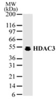
- Submitted by
- Invitrogen Antibodies (provider)
- Main image

- Experimental details
- Western blot analysis of HDAC3 in HeLa cell lysate probed with a HDAC3 polyclonal antibody (Product # PA1-41039).

- Submitted by
- Invitrogen Antibodies (provider)
- Main image

- Experimental details
- Western blot analysis of HDAC3 in HeLa cell lysate. Sample was incubated in HDAC3 polyclonal antibody (Product # PA1-41039).
- Submitted by
- Invitrogen Antibodies (provider)
- Main image

- Experimental details
- Western blot analysis of HDAC3 in 1.0 mg/mL HeLa lysate. Samples were incubated in HDAC3 polyclonal (Product # PA1-41039). This experiment was performed under reducing conditions using the 12-230 kDa separation system.

Supportive validation
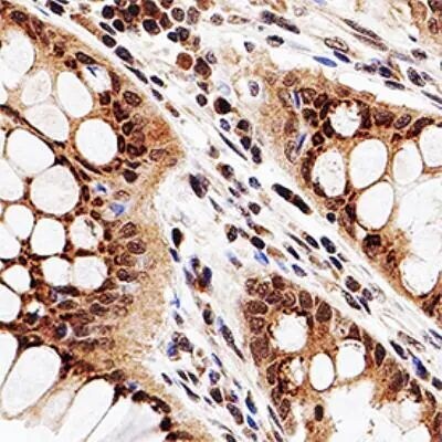
- Submitted by
- Invitrogen Antibodies (provider)
- Main image
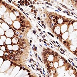
- Experimental details
- Immunohistochemical analysis of HDAC3 in human colon cancer. Samples were incubated in HDAC3 polyclonal antibody (Product # PA1-41039) using a dilution of 1:50. Bond Rx autostainer (Leica Biosystems). The assay involved 20 minutes of heat induced antigen retrieval (HIER) using 10mM sodium citrate buffer (pH 6.0) and endogenous peroxidase quenching with peroxide block. The sections were incubated with primary antibody for 30 minutes and Bond Polymer Refine Detection (Leica Biosystems) with DAB was used for signal development followed by counterstaining with hematoxylin. Whole slide scanning and capturing of representative images was performed using Aperio AT2 (Leica Biosystems). Nuclear staining of HDAC3 was observed. Staining was performed by Histowiz.