 Explore
 Validate
 Learn
Learn Western blot
Antibody data
- Antibody Data
- Antigen structure
- References [5]
- Comments [0]
- Validations
- Western blot [1]
- Immunocytochemistry [1]
- Immunohistochemistry [1]
- Chromatin Immunoprecipitation [1]
Submit
Validation data
Reference
Comment
Report error
- Product number
- PA1-862 - Provider product page

- Provider
- Invitrogen Antibodies
- Product name
- HDAC3 Polyclonal Antibody
- Antibody type
- Polyclonal
- Antigen
- Synthetic peptide
- Description
- PA1-862 detects histone deacetylase 3 (HDAC3) from canine, hamster, human and rat tissues and cells.
- Concentration
- 1 mg/mL
Submitted references Regulated clearance of histone deacetylase 3 protects independent formation of nuclear receptor corepressor complexes.
The transcriptional repressor Sp3 is associated with CK2-phosphorylated histone deacetylase 2.
Effect of estradiol on histone acetylation dynamics in human breast cancer cells.
Characterization of a human RPD3 ortholog, HDAC3.
Differential display cloning of a novel human histone deacetylase (HDAC3) cDNA from PHA-activated immune cells.
Guo C, Gow CH, Li Y, Gardner A, Khan S, Zhang J
The Journal of biological chemistry 2012 Apr 6;287(15):12111-20
The Journal of biological chemistry 2012 Apr 6;287(15):12111-20
The transcriptional repressor Sp3 is associated with CK2-phosphorylated histone deacetylase 2.
Sun JM, Chen HY, Moniwa M, Litchfield DW, Seto E, Davie JR
The Journal of biological chemistry 2002 Sep 27;277(39):35783-6
The Journal of biological chemistry 2002 Sep 27;277(39):35783-6
Effect of estradiol on histone acetylation dynamics in human breast cancer cells.
Sun JM, Chen HY, Davie JR
The Journal of biological chemistry 2001 Dec 28;276(52):49435-42
The Journal of biological chemistry 2001 Dec 28;276(52):49435-42
Characterization of a human RPD3 ortholog, HDAC3.
Emiliani S, Fischle W, Van Lint C, Al-Abed Y, Verdin E
Proceedings of the National Academy of Sciences of the United States of America 1998 Mar 17;95(6):2795-800
Proceedings of the National Academy of Sciences of the United States of America 1998 Mar 17;95(6):2795-800
Differential display cloning of a novel human histone deacetylase (HDAC3) cDNA from PHA-activated immune cells.
Dangond F, Hafler DA, Tong JK, Randall J, Kojima R, Utku N, Gullans SR
Biochemical and biophysical research communications 1998 Jan 26;242(3):648-52
Biochemical and biophysical research communications 1998 Jan 26;242(3):648-52
No comments: Submit comment
Supportive validation
- Submitted by
- Invitrogen Antibodies (provider)
- Main image
- Experimental details
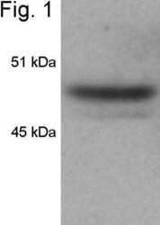
- Western blot of HDAC3 from HeLa cell lysate using Product # PA1-862.

Supportive validation
- Submitted by
- Invitrogen Antibodies (provider)
- Main image

- Experimental details
- Immunofluorescent analysis of HDAC3 in HeLa Cells. Cells were grown on chamber slides and fixed with formaldehyde prior to staining. Cells were probed without (control) or with a HDAC3 polyclonal antibody (Product # PA1-862) at a dilution of 1:200 overnight at 4 C, washed with PBS and incubated with a DyLight-488 conjugated secondary antibody (Product # 35552). HDAC3 staining (green), F-Actin staining with Phalloidin (red) and nuclei with DAPI (blue) is shown. Images were taken at 60X magnification.

Supportive validation
- Submitted by
- Invitrogen Antibodies (provider)
- Main image

- Experimental details
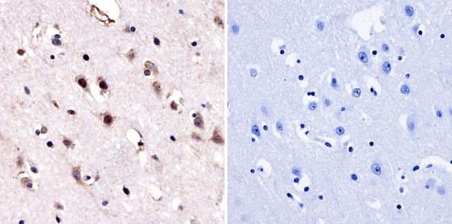
- Immunohistochemistry was performed on normal biopsies of deparaffinized human brain tissue. To expose target proteins, heat induced antigen retrieval was performed using 10mM sodium citrate (pH6.0) buffer, microwaved for 8-15 minutes. Following antigen retrieval tissues were blocked in 3% BSA-PBS for 30 minutes at room temperature. Tissues were then probed at a dilution of 1:50 with a Rabbit Polyclonal Antibody recognizing HDAC3 (Product # PA1-862) or without primary antibody (negative control) overnight at 4°C in a humidified chamber. Tissues were washed extensively with PBST and endogenous peroxidase activity was quenched with a peroxidase suppressor. Detection was performed using a biotin-conjugated secondary antibody and SA-HRP, followed by colorimetric detection using DAB. Tissues were counterstained with hematoxylin and prepped for mounting.
Supportive validation
- Submitted by
- Invitrogen Antibodies (provider)
- Main image

- Experimental details
- Chromatin immunoprecipitation analysis of HDAC3 was performed using cross-linked chromatin from 1x10^6 HCT116 colon carcinoma cells treated with serum for 0, 15, and 30 minutes. Immunoprecipitation was performed using a multiplex microplate Matrix ChIP assay (see reference for Matrix ChIP protocol: http://www.ncbi.nlm.nih.gov/pubmed/22098709) with 1.0 µL/100 µL well volume of an HDAC3 polyclonal antibody (Product # PA1-862). Chromatin aliquots from ~1x10^5 cells were used per ChIP pull-down. Quantitative PCR data were done in quadruplicate using 1 µL of eluted DNA in 2 µL SYBR real-time PCR reactions containing primers to amplify -15kb upstream of the Egr1 gene or exon-1 or exon-2 of Egr1. PCR calibration curves were generated for each primer pair from a dilution series of sheared total genomic DNA. Quantitation of immunoprecipitated chromatin is presented as signal relative to the total amount of input chromatin. Results represent the mean +/- SEM for three experiments. A schematic representation of the Egr-1 locus is shown above the data where boxes represent exons (black boxes = translated regions, white boxes = untranslated regions), the zigzag line represents an intron, and the straight line represents upstream sequence. Regions amplified by Egr-1 primers are represented by black bars. Data courtesy of the Innovators Program.
