Explore
 Validate
 Learn
Learn Western blot
 Immunohistochemistry
Antibody data
- Antibody Data
- Antigen structure
- References [2]
- Comments [0]
- Validations
- Immunohistochemistry [1]
Submit
Validation data
Reference
Comment
Report error
- Product number
- PA1600-1 - Provider product page

- Provider
- Boster Biological Technology
- Product name
- Anti-Histone deacetylase 3 HDAC3 Antibody
- Antibody type
- Polyclonal
- Description
- Polyclonal antibody for HDAC3 detection. Host: Rabbit.Size: 100μg/vial. Tested applications: WB, IHC-P, IHC-F, ICC/IF, FCM. Reactive species: Human. HDAC3 information: Molecular Weight: 48848 MW; Subcellular Localization: Nucleus . Cytoplasm . Cytoplasm, cytosol . Colocalizes with XBP1 and AKT1 in the cytoplasm (PubMed:25190803). Predominantly expressed in the nucleus in the presence of CCAR2; Tissue Specificity: Widely expressed.
- Reactivity
- Human, Mouse, Rat
- Host
- Rabbit
- Vial size
- 100μg/vial
- Concentration
- Add 0.2ml of distilled water will yield a concentration of 500ug/ml.
- Storage
- At -20°C for one year. After reconstitution, at 4°C for one month. It can also be aliquoted and stored frozen at -20°C for a longer time. Avoid repeated freezing and thawing.
- Handling
- Add 0.2ml of distilled water will yield a concentration of 500ug/ml.
Submitted references The key role of macrophage depolarization in the treatment of COPD with ergosterol both in vitro and in vivo.
Interleukin-18 Down-Regulates Multidrug Resistance-Associated Protein 2 Expression through Farnesoid X Receptor Associated with Nuclear Factor Kappa B and Yin Yang 1 in Human Hepatoma HepG2 Cells.
Sun X, Liu Y, Feng X, Li C, Li S, Zhao Z
International immunopharmacology 2020 Feb;79:106086
International immunopharmacology 2020 Feb;79:106086
Interleukin-18 Down-Regulates Multidrug Resistance-Associated Protein 2 Expression through Farnesoid X Receptor Associated with Nuclear Factor Kappa B and Yin Yang 1 in Human Hepatoma HepG2 Cells.
Liu XC, Lian W, Zhang LJ, Feng XC, Gao Y, Li SX, Liu C, Cheng Y, Yang L, Wang XJ, Chen L, Wang RQ, Chai J, Chen WS
PloS one 2015;10(8):e0136215
PloS one 2015;10(8):e0136215
No comments: Submit comment
Supportive validation
- Submitted by
- Boster Biological Technology (provider)
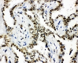
- Main image
- Experimental details
- IHC analysis of HDAC3 using anti-HDAC3 antibody (PA1600-1). HDAC3 was detected in paraffin-embedded section of human intestinal cancer tissues. Heat mediated antigen retrieval was performed in citrate buffer (pH6, epitope retrieval solution) for 20 mins. The tissue section was blocked with 10% goat serum. The tissue section was then incubated with 1μg/ml rabbit anti-HDAC3 Antibody (PA1600-1) overnight at 4°C. Biotinylated goat anti-rabbit IgG was used as secondary antibody and incubated for 30 minutes at 37°C. The tissue section was developed using Strepavidin-Biotin-Complex (SABC)(Catalog # SA1022) with DAB as the chromogen.
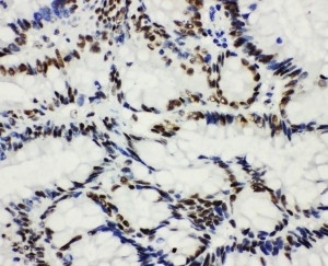
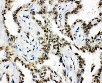
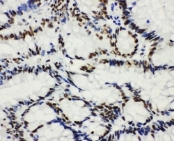
- Additional image