 Explore
 Validate
 Learn
Learn Western blot
 Immunoprecipitation
Antibody data
- Antibody Data
- Antigen structure
- References [0]
- Comments [0]
- Validations
- Western blot [2]
- Immunocytochemistry [1]
- Immunohistochemistry [1]
- Other assay [1]
Submit
Validation data
Reference
Comment
Report error
- Product number
- PA5-96776 - Provider product page

- Provider
- Invitrogen Antibodies
- Product name
- MAP2K4 Polyclonal Antibody
- Antibody type
- Polyclonal
- Antigen
- Synthetic peptide
- Description
- Positive Samples: BT-474, MCF7, SH-SY5Y, Rat skeletal muscle ; Cellular Location: Cytoplasm, Nucleus
- Reactivity
- Human, Mouse, Rat
- Host
- Rabbit
- Isotype
- IgG
- Vial size
- 100 µL
- Concentration
- 1.84 mg/mL
- Storage
- -20° C, Avoid Freeze/Thaw Cycles
No comments: Submit comment
Supportive validation
- Submitted by
- Invitrogen Antibodies (provider)
- Main image

- Experimental details
- Western blot was performed using MAP2K4 Polyclonal Antibody (Product # PA5-96776) and a 44 kDa band corresponding to MAP2K4 protein was observed across across all the cell lines. Whole cell extracts (30 µg lysate) of HeLa (Lane 1), HEK-293 (Lane 2), NIH/3T3 (Lane 3) were electrophoresed using NuPAGE™ 4-12% Bis-Tris Protein Gel (Product # NP0321BOX), 10 well. Resolved proteins were then transferred onto a nitrocellulose membrane (Product # IB23001) by iBlot® 2 Dry Blotting System (Product # IB21001). The blot was probed with the primary antibody (1:1000 dilution) and detected by chemiluminescence with Goat anti-Rabbit IgG (H+L) Superclonal™ Recombinant Secondary Antibody, HRP (Product # A27036, 1:10,000 dilution) using the iBright™ FL1500 Imaging System (Product # A44115). Chemiluminescent detection was performed using SuperSignal™ West Pico PLUS Chemiluminescent Substrate (Product # 34580).

- Submitted by
- Invitrogen Antibodies (provider)
- Main image

- Experimental details
- Western Blot analysis of SEK1 in extracts of various cell lines using SEK1 Polyclonal Antibody (Product # PA5-96776) at a dilution of 1:1000. A HRP Goat Anti-Rabbit IgG (H+L) secondary antibody was used at a dilution of 1:10,000. Lysates/proteins: 25 µg per lane. Blocking buffer: 3% nonfat dry milk in TBST.

Supportive validation
- Submitted by
- Invitrogen Antibodies (provider)
- Main image
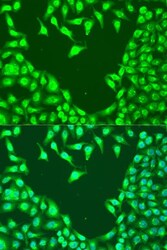
- Experimental details
- Immunocytochemistry-Immunofluorescence analysis of SEK1 was performed in U2OS cells using SEK1 Polyclonal Antibody (Product # PA5-96776) at a dilution of 1:100. Blue: DAPI for nuclear staining.

Supportive validation
- Submitted by
- Invitrogen Antibodies (provider)
- Main image
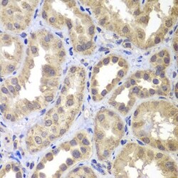
- Experimental details
- Immunohistochemistry analysis of SEK1 in paraffin-embedded human kidney using SEK1 Polyclonal Antibody (Product # PA5-96776) at a dilution of 1:100.

Supportive validation
- Submitted by
- Invitrogen Antibodies (provider)
- Main image

- Experimental details
- Immunoprecipitation analysis of SEK1 was performed in 200 µg extracts of MCF-7 cells using SEK1 Polyclonal Antibody (Product # PA5-96776). Western blot was performed from the immunoprecipitate using SEK1 Polyclonal Antibody at a dilution of 1:1000.
