Explore
 Validate
 Learn
Learn Western blot
 Immunocytochemistry
 Immunohistochemistry
Antibody data
- Antibody Data
- Antigen structure
- References [4]
- Comments [0]
- Validations
- Western blot [1]
- Immunocytochemistry [1]
Submit
Validation data
Reference
Comment
Report error
- Product number
- HPA022434 - Provider product page

- Provider
- Atlas Antibodies
- Proper citation
- Atlas Antibodies Cat#HPA022434, RRID:AB_1844513
- Product name
- Anti-ACLY
- Antibody type
- Polyclonal
- Description
- Polyclonal Antibody against Human ACLY, Gene description: ATP citrate lyase, Alternative Gene Names: ACL, ATPCL, CLATP, Validated applications: WB, IHC, ICC, Uniprot ID: P53396, Storage: Store at +4°C for short term storage. Long time storage is recommended at -20°C.
- Reactivity
- Human, Mouse, Rat
- Host
- Rabbit
- Conjugate
- Unconjugated
- Isotype
- IgG
- Vial size
- 100 µl
- Concentration
- 0.1 mg/ml
- Storage
- Store at +4°C for short term storage. Long time storage is recommended at -20°C.
- Handling
- The antibody solution should be gently mixed before use.
Submitted references Cytoskeletal association of ATP citrate lyase controls the mechanodynamics of macropinocytosis
ONECUT2 facilitates hepatocellular carcinoma metastasis by transcriptionally upregulating FGF2 and ACLY.
Autophagy regulates fatty acid availability for oxidative phosphorylation through mitochondria-endoplasmic reticulum contact sites.
ACLY (ATP Citrate Lyase) Mediates Radioresistance in Head and Neck Squamous Cell Carcinomas and is a Novel Predictive Radiotherapy Biomarker
Puccini J, Wei J, Tong L, Bar-Sagi D
Proceedings of the National Academy of Sciences 2023;120(8)
Proceedings of the National Academy of Sciences 2023;120(8)
ONECUT2 facilitates hepatocellular carcinoma metastasis by transcriptionally upregulating FGF2 and ACLY.
Liu D, Zhang T, Chen X, Zhang B, Wang Y, Xie M, Ji X, Sun M, Huang W, Xia L
Cell death & disease 2021 Nov 27;12(12):1113
Cell death & disease 2021 Nov 27;12(12):1113
Autophagy regulates fatty acid availability for oxidative phosphorylation through mitochondria-endoplasmic reticulum contact sites.
Bosc C, Broin N, Fanjul M, Saland E, Farge T, Courdy C, Batut A, Masoud R, Larrue C, Skuli S, Espagnolle N, Pagès JC, Carrier A, Bost F, Bertrand-Michel J, Tamburini J, Récher C, Bertoli S, Mansat-De Mas V, Manenti S, Sarry JE, Joffre C
Nature communications 2020 Aug 13;11(1):4056
Nature communications 2020 Aug 13;11(1):4056
ACLY (ATP Citrate Lyase) Mediates Radioresistance in Head and Neck Squamous Cell Carcinomas and is a Novel Predictive Radiotherapy Biomarker
Göttgens E, van den Heuvel C, de Jong M, Kaanders J, Leenders W, Ansems M, Bussink J, Span P
Cancers 2019;11(12):1971
Cancers 2019;11(12):1971
No comments: Submit comment
Enhanced validation
- Submitted by
- Atlas Antibodies (provider)
- Enhanced method
- Genetic validation
- Main image
- Experimental details
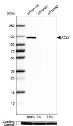
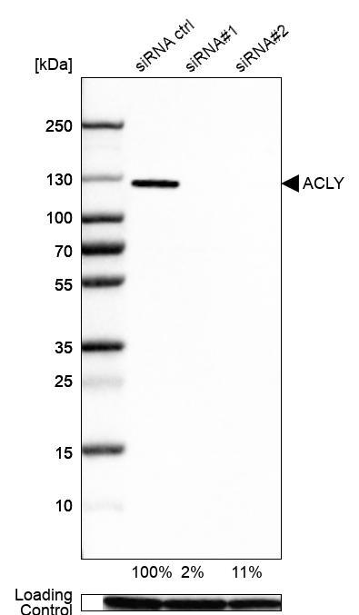
- Western blot analysis in A-549 cells transfected with control siRNA, target specific siRNA probe #1 and #2, using Anti-ACLY antibody. Remaining relative intensity is presented. Loading control: Anti-GAPDH.
- Sample type
- Human
- Protocol
- Protocol
Supportive validation
- Submitted by
- Atlas Antibodies (provider)
- Main image
- Experimental details
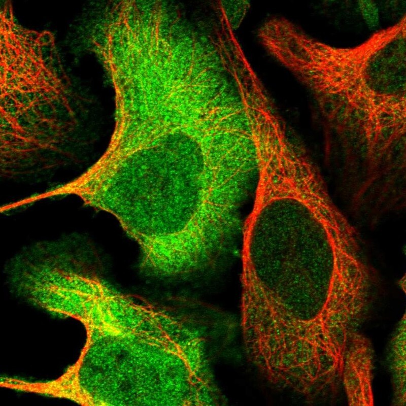
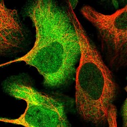
- Immunofluorescent staining of human cell line U-2 OS shows localization to nucleoplasm, plasma membrane & cytosol.
- Sample type
- Human