 Explore
 Validate
 Learn
Learn Western blot
Antibody data
- Antibody Data
- Antigen structure
- References [4]
- Comments [0]
- Validations
- Western blot [2]
- Immunocytochemistry [1]
Submit
Validation data
Reference
Comment
Report error
- Product number
- 702731 - Provider product page

- Provider
- Invitrogen Antibodies
- Product name
- ATP Citrate Lyase Recombinant Rabbit Monoclonal Antibody (4H6L2)
- Antibody type
- Monoclonal
- Antigen
- Other
- Reactivity
- Human
- Host
- Rabbit
- Isotype
- IgG
- Antibody clone number
- 4H6L2
- Vial size
- 100 µg
- Concentration
- 0.5 mg/mL
- Storage
- Store at 4°C short term. For long term storage, store at -20°C, avoiding freeze/thaw cycles.
Submitted references Glutamate triggers the expression of functional ionotropic and metabotropic glutamate receptors in mast cells.
Distinct B cell subsets in Peyer's patches convey probiotic effects by Limosilactobacillus reuteri.
Transcription factor FOXP2 is a flow-induced regulator of collecting lymphatic vessels.
Transcriptome profiling data reveals ubiquitin-specific peptidase 9 knockdown effects.
Alim MA, Grujic M, Ackerman PW, Kristiansson P, Eliasson P, Peterson M, Pejler G
Cellular & molecular immunology 2021 Oct;18(10):2383-2392
Cellular & molecular immunology 2021 Oct;18(10):2383-2392
Distinct B cell subsets in Peyer's patches convey probiotic effects by Limosilactobacillus reuteri.
Liu HY, Giraud A, Seignez C, Ahl D, Guo F, Sedin J, Walden T, Oh JH, van Pijkeren JP, Holm L, Roos S, Bertilsson S, Phillipson M
Microbiome 2021 Oct 3;9(1):198
Microbiome 2021 Oct 3;9(1):198
Transcription factor FOXP2 is a flow-induced regulator of collecting lymphatic vessels.
Hernández Vásquez MN, Ulvmar MH, González-Loyola A, Kritikos I, Sun Y, He L, Halin C, Petrova TV, Mäkinen T
The EMBO journal 2021 Jun 15;40(12):e107192
The EMBO journal 2021 Jun 15;40(12):e107192
Transcriptome profiling data reveals ubiquitin-specific peptidase 9 knockdown effects.
Glaab E, Antony P, Köglsberger S, Forster JI, Cordero-Maldonado ML, Crawford A, Garcia P, Buttini M
Data in brief 2019 Aug;25:104130
Data in brief 2019 Aug;25:104130
No comments: Submit comment
Supportive validation
- Submitted by
- Invitrogen Antibodies (provider)
- Main image

- Experimental details
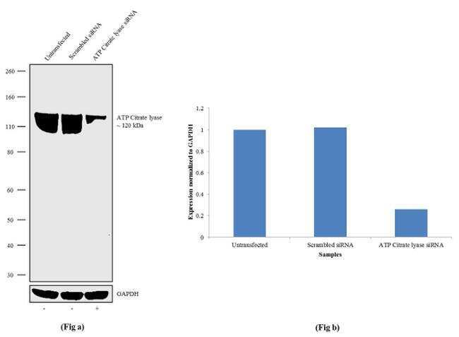
- Knockdown of ATP Citrate lyase was achieved by transfecting A549 cells with ATP Citrate lyase specific validated siRNA (Silencer® select Product # s916). Western blot analysis (Fig a) was performed using Whole cell extracts from the ATP Citrate lyase knock down cells (Lane 3), non-specific scrambled siRNA transfected cells (Lane 2) and untransfected cells (Lane 1). The blots were probed with Anti-ATP Citrate lyase Recombinant Rabbit Monoclonal Antibody (Product # 702731, 1-3 µg/mL) and Goat anti-Rabbit IgG (H+L) Superclonal™ Secondary Antibody, HRP conjugate (Product # A27036, 0.25 µg/mL, 1:4000 dilution). Densitometric analysis of this Western blot is shown in histogram (Fig b). Loss of signal upon siRNA mediated knock down confirms that antibody is specific to ATP Citrate lyase.
- Submitted by
- Invitrogen Antibodies (provider)
- Main image

- Experimental details
- Western blot analysis was performed on Whole cell extracts (30 µg lysate) of A549 (Lane 1), HeLa (Lane 2), NCI-H1975 (Lane 3), PC-3 (Lane 4), SK-OV-3 (Lane 5), A-431 (Lane 6), and Jurkat (Lane7). The blots were probed with Anti-ATP Citrate lyase Recombinant Rabbit Monoclonal Antibody (Product # 702731, 2.5 µg/mL) and detected by chemiluminescence using Goat anti-Rabbit IgG (H+L) Superclonal™ Secondary Antibody, HRP conjugate (Product # A27036, 0.25 µg/mL, 1:4000 dilution). A 120 kDa band corresponding to ATP Citrate lyase was observed across the cell lines tested. Known quantity of protein samples were electrophoresed using Novex®NuPAGE®4-12% Bis-Tris gel (Product # NP0322BOX), XCell SureLock™ Electrophoresis System (Product # EI0002) and Novex® Sharp Pre-Stained Protein Standard (Product # LC5800). Resolved proteins were then transferred onto a nitrocellulose membrane with iBlot® Dry Blotting System (Product # IB21001). The membrane was probed with the relevant primary and secondary Antibody following blocking with 5% skimmed milk. Chemiluminescent detection was performed using Pierce™ ECL Western blotting Substrate (Product # 32106).

Supportive validation
- Submitted by
- Invitrogen Antibodies (provider)
- Main image

- Experimental details
- For immunofluorescence analysis, A549 cells were fixed and permeabilized for detection of endogenous ATP Citrate Lyase using Anti- ATP Citrate Lyase Recombinant Rabbit Monoclonal Antibody (Product # 702731, 5 µg/mL) and labeled with Goat anti-Rabbit IgG (H+L) Superclonal™ Secondary Antibody, Alexa Fluor® 488 conjugate (Product # A27034, 1:2000). Panel a) shows representative cells that were stained for detection and localization of ATP Citrate Lyase protein (green), Panel b) is stained for nuclei (blue) using SlowFade® Gold Antifade Mountant with DAPI (Product # S36938). Panel c) represents cytoskeletal F-actin staining using Rhodamine Phalloidin (Product # R415, 1:300). Panel d) is a composite image of Panels a, b and c clearly demonstrating cytoplasmic localization of ATP Citrate Lyase. Panel e) represents control cells with no primary antibody to assess background. The images were captured at 60X magnification.
