Explore
 Validate
 Learn
Learn Western blot
Antibody data
- Antibody Data
- Antigen structure
- References [0]
- Comments [0]
- Validations
- Western blot [4]
- Immunocytochemistry [1]
- Immunohistochemistry [16]
Submit
Validation data
Reference
Comment
Report error
- Product number
- MA5-25067 - Provider product page

- Provider
- Invitrogen Antibodies
- Product name
- ATP Citrate Lyase Monoclonal Antibody (OTI3G8)
- Antibody type
- Monoclonal
- Antigen
- Recombinant full-length protein
- Reactivity
- Human
- Host
- Mouse
- Isotype
- IgG
- Antibody clone number
- OTI3G8
- Vial size
- 100 µL
- Concentration
- 0.58 mg/mL
- Storage
- -20° C, Avoid Freeze/Thaw Cycles
No comments: Submit comment
Supportive validation
- Submitted by
- Invitrogen Antibodies (provider)
- Main image
- Experimental details
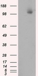
- Western blot analysis of ACLY in HEK293T cells in untransfected (Left lane) and transfected (Right lane) samples using 5 µg per lane. The samples were separated by SDS-PAGE and probed with ACLY (Product # MA5-25067) monoclonal antibody.
- Submitted by
- Invitrogen Antibodies (provider)
- Main image
- Experimental details
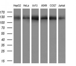
- Western blot analysis of ACLY in HepG2, HeLa, STV2, A549, COS7, Jurkat cells using 10 µg per lane. Samples were probed with ACLY (Product # MA5-25067) monoclonal antibody at a dilution of 1:200.

- Submitted by
- Invitrogen Antibodies (provider)
- Main image

- Experimental details
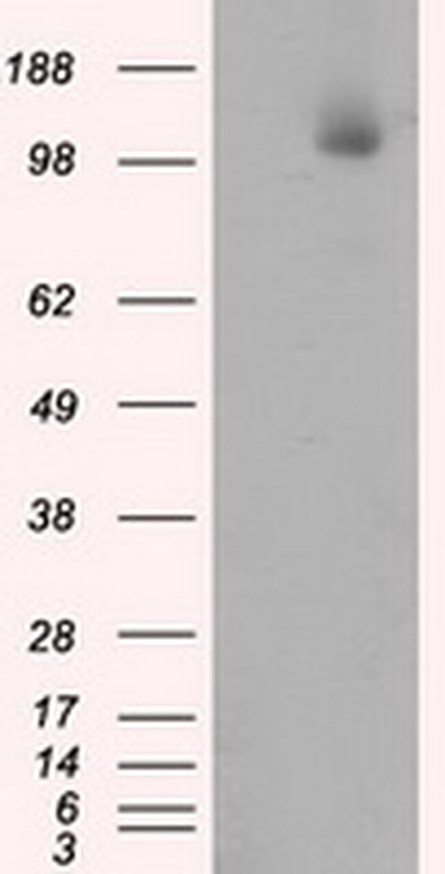
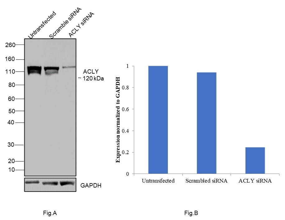
- Knockdown of ATP Citrate Lyase was achieved by transfecting A549 with ATP Citrate Lyase specific siRNAs (Silencer® select Product # S915, S916). Western blot analysis (Fig. a) was performed using Whole cell extracts from the ATP Citrate Lyase untransfected cells (lane 1), non-targeting scrambled siRNA transfected cells (lane 2) and knockdown cells (lane 3). The blot was probed with ATP Citrate Lyase Monoclonal Antibody (OTI3G8) (Product # MA5-25067, 1:1000 dilution ) and Goat anti-Mouse IgG (H+L) Superclonal™ Recombinant Secondary Antibody, HRP (Product # A28177, 1:4000 dilution). Densitometric analysis of this western blot is shown in histogram (Fig. b). Decrease in signal upon siRNA mediated knock down confirms that antibody is specific to ATP Citrate Lyase.
- Submitted by
- Invitrogen Antibodies (provider)
- Main image
- Experimental details
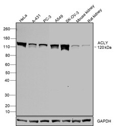
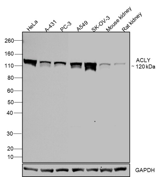
- Western blot was performed using Anti-ATP Citrate Lyase Monoclonal Antibody (OTI3G8)(Product # MA5-25067) and a 120kDa band corresponding to ATP Citrate Lyase was observed across cell lines and tissues tested. Whole cell extracts (30 µg lysate) of HeLa (Lane 1), A-431 (Lane 2), PC-3 (Lane 3), A549 (Lane 4), SK-O-V3 (Lane 5), Mouse Kidney (Lane 6), Rat Kidney (Lane 7) were electrophoresed using NuPAGE™ 4-12% Bis-Tris Protein Gel (Product # NP0321BOX). Resolved proteins were then transferred onto a Nitrocellulose membrane (Product # IB23001) by iBlot® 2 Dry Blotting System (Product # IB21001). The blot was probed with the primary antibody (1:1000 Dilution) and detected by chemiluminescence with Goat anti-Mouse IgG (H+L) Superclonal™ Recombinant Secondary Antibody, HRP (Product # A28177,1:4000 dilution) using the iBright FL 1000 (Product # A32752). Chemiluminescent detection was performed using Novex® ECL Chemiluminescent Substrate Reagent Kit (Product # WP20005).
Supportive validation
- Submitted by
- Invitrogen Antibodies (provider)
- Main image

- Experimental details
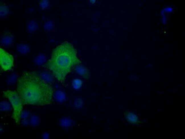
- Immunofluorescent analysis of ACLY in COS7 cells. Cells were transfected with a plasmid overexpressing ACLY and probed with a ACLY monoclonal antibody (Product # MA5-25067).
Supportive validation
- Submitted by
- Invitrogen Antibodies (provider)
- Main image
- Experimental details
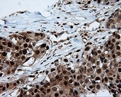
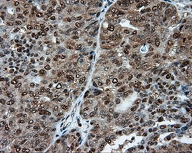
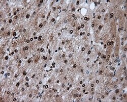
- Immunohistochemistry was performed on paraffin-embedded adenocarcinoma of breast tissue. To expose target proteins, 10mM citric buffer, pH6.0, 100°C for 10min was used. Following antigen retrieval, tissues were probed with a ACLY monoclonal antibody (Product # MA5-25067) at a dilution of 1:50.

- Submitted by
- Invitrogen Antibodies (provider)
- Main image
- Experimental details
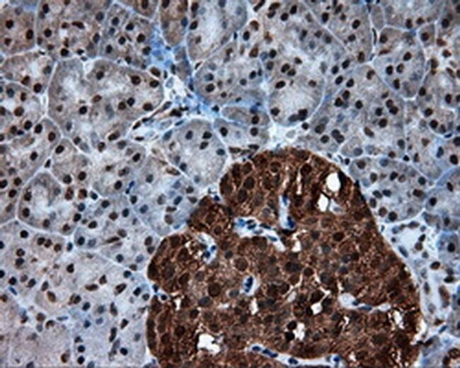
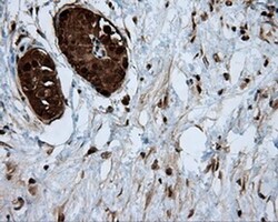
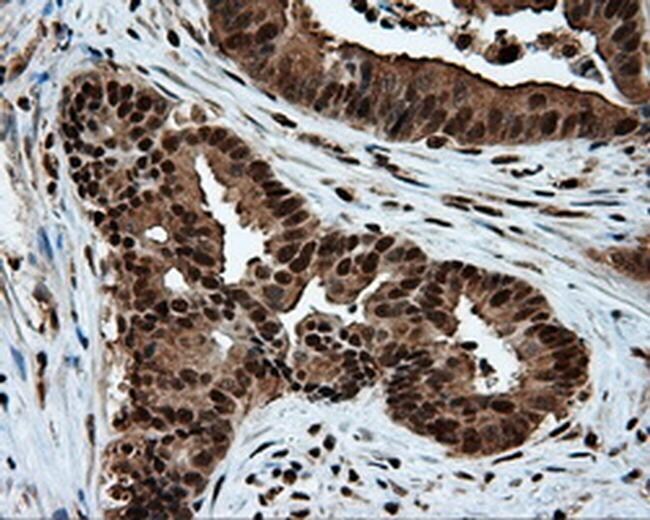
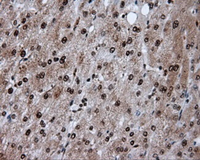
- Immunohistochemistry was performed on paraffin-embedded carcinoma of pancreas tissue. To expose target proteins, 10mM citric buffer, pH6.0, 100°C for 10min was used. Following antigen retrieval, tissues were probed with a ACLY monoclonal antibody (Product # MA5-25067) at a dilution of 1:50.

- Submitted by
- Invitrogen Antibodies (provider)
- Main image
- Experimental details
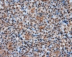
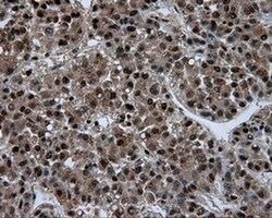
- Immunohistochemistry was performed on paraffin-embedded carcinoma of thyroid tissue. To expose target proteins, 10mM citric buffer, pH6.0, 100°C for 10min was used. Following antigen retrieval, tissues were probed with a ACLY monoclonal antibody (Product # MA5-25067) at a dilution of 1:50.

- Submitted by
- Invitrogen Antibodies (provider)
- Main image

- Experimental details
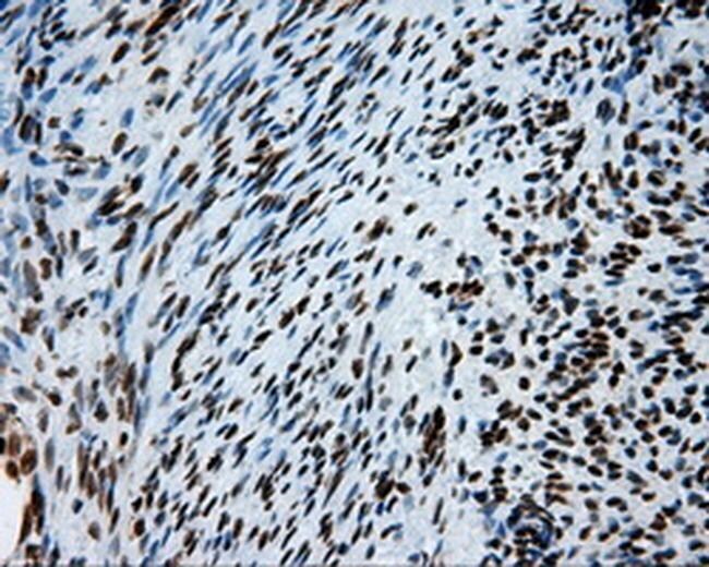
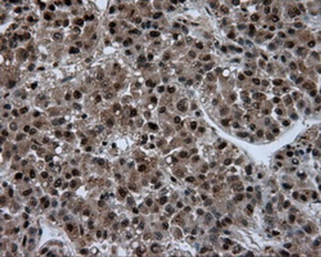
- Immunohistochemistry was performed on paraffin-embedded endometrium tissue. To expose target proteins, 10mM citric buffer, pH6.0, 100°C for 10min was used. Following antigen retrieval, tissues were probed with a ACLY monoclonal antibody (Product # MA5-25067) at a dilution of 1:50.
- Submitted by
- Invitrogen Antibodies (provider)
- Main image

- Experimental details
- Immunohistochemistry was performed on paraffin-embedded adenocarcinoma of endometrium tissue. To expose target proteins, 10mM citric buffer, pH6.0, 100°C for 10min was used. Following antigen retrieval, tissues were probed with a ACLY monoclonal antibody (Product # MA5-25067) at a dilution of 1:50.
- Submitted by
- Invitrogen Antibodies (provider)
- Main image

- Experimental details
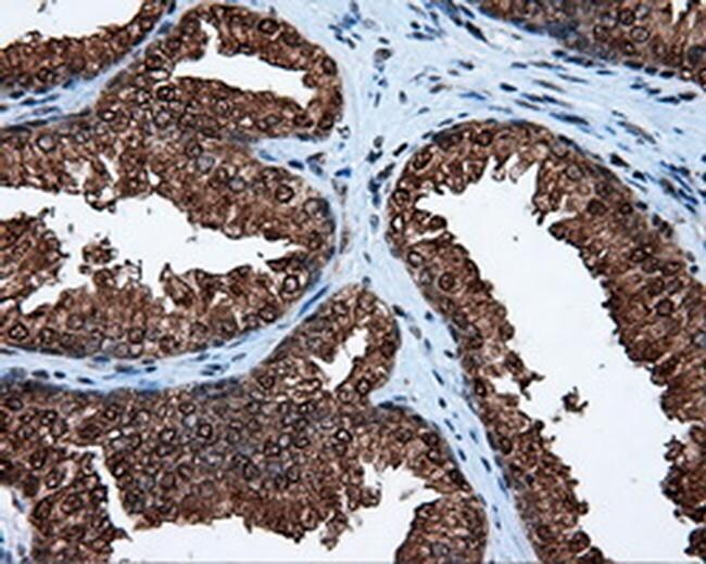
- Immunohistochemistry was performed on paraffin-embedded prostate tissue. To expose target proteins, 10mM citric buffer, pH6.0, 100°C for 10min was used. Following antigen retrieval, tissues were probed with a ACLY monoclonal antibody (Product # MA5-25067) at a dilution of 1:50.
- Submitted by
- Invitrogen Antibodies (provider)
- Main image

- Experimental details
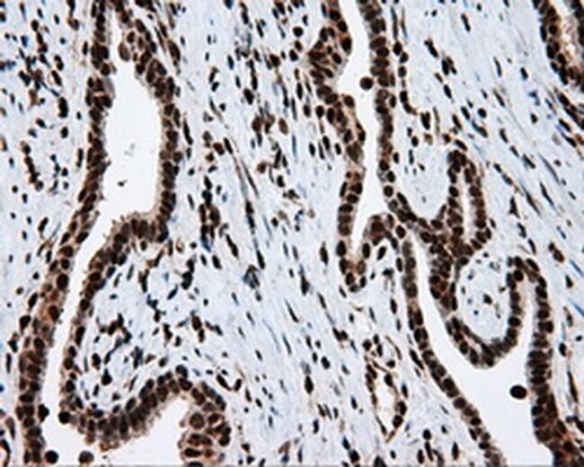
- Immunohistochemistry was performed on paraffin-embedded carcinoma of prostate tissue. To expose target proteins, 10mM citric buffer, pH6.0, 100°C for 10min was used. Following antigen retrieval, tissues were probed with a ACLY monoclonal antibody (Product # MA5-25067) at a dilution of 1:50.
- Submitted by
- Invitrogen Antibodies (provider)
- Main image
- Experimental details
- Immunohistochemistry was performed on paraffin-embedded bladder tissue. To expose target proteins, 10mM citric buffer, pH6.0, 100°C for 10min was used. Following antigen retrieval, tissues were probed with a ACLY monoclonal antibody (Product # MA5-25067) at a dilution of 1:50.

- Submitted by
- Invitrogen Antibodies (provider)
- Main image
- Experimental details
- Immunohistochemistry was performed on paraffin-embedded colon tissue. To expose target proteins, 10mM citric buffer, pH6.0, 100°C for 10min was used. Following antigen retrieval, tissues were probed with a ACLY monoclonal antibody (Product # MA5-25067) at a dilution of 1:50.
- Submitted by
- Invitrogen Antibodies (provider)
- Main image

- Experimental details
- Immunohistochemistry was performed on paraffin-embedded adenocarcinoma of colon tissue. To expose target proteins, 10mM citric buffer, pH6.0, 100°C for 10min was used. Following antigen retrieval, tissues were probed with a ACLY monoclonal antibody (Product # MA5-25067) at a dilution of 1:50.
- Submitted by
- Invitrogen Antibodies (provider)
- Main image
- Experimental details
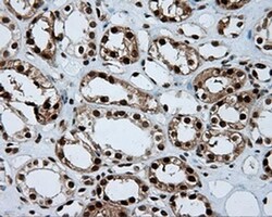
- Immunohistochemistry was performed on paraffin-embedded kidney tissue. To expose target proteins, 10mM citric buffer, pH6.0, 100°C for 10min was used. Following antigen retrieval, tissues were probed with a ACLY monoclonal antibody (Product # MA5-25067) at a dilution of 1:50.

- Submitted by
- Invitrogen Antibodies (provider)
- Main image
- Experimental details
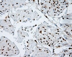
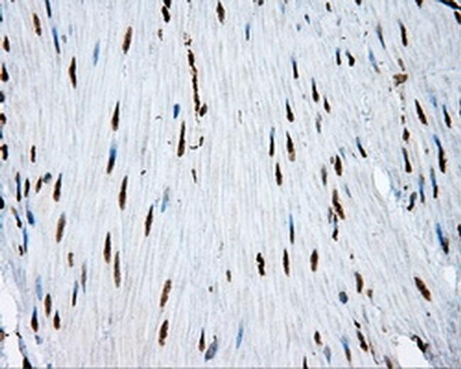
- Immunohistochemistry was performed on paraffin-embedded liver tissue. To expose target proteins, 10mM citric buffer, pH6.0, 100°C for 10min was used. Following antigen retrieval, tissues were probed with a ACLY monoclonal antibody (Product # MA5-25067) at a dilution of 1:50.
- Submitted by
- Invitrogen Antibodies (provider)
- Main image
- Experimental details
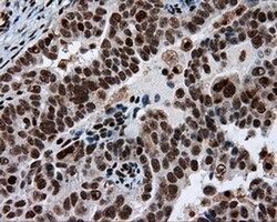
- Immunohistochemistry was performed on paraffin-embedded carcinoma of liver tissue. To expose target proteins, 10mM citric buffer, pH6.0, 100°C for 10min was used. Following antigen retrieval, tissues were probed with a ACLY monoclonal antibody (Product # MA5-25067) at a dilution of 1:50.
- Submitted by
- Invitrogen Antibodies (provider)
- Main image
- Experimental details
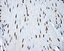
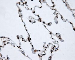
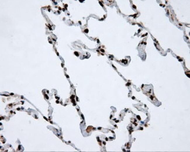
- Immunohistochemistry was performed on paraffin-embedded lung tissue. To expose target proteins, 10mM citric buffer, pH6.0, 100°C for 10min was used. Following antigen retrieval, tissues were probed with a ACLY monoclonal antibody (Product # MA5-25067) at a dilution of 1:50.
- Submitted by
- Invitrogen Antibodies (provider)
- Main image
- Experimental details
- Immunohistochemistry was performed on paraffin-embedded adenocarcinoma of ovary tissue. To expose target proteins, 10mM citric buffer, pH6.0, 100°C for 10min was used. Following antigen retrieval, tissues were probed with a ACLY monoclonal antibody (Product # MA5-25067) at a dilution of 1:50.

- Submitted by
- Invitrogen Antibodies (provider)
- Main image

- Experimental details
- Immunohistochemistry was performed on paraffin-embedded pancreas tissue. To expose target proteins, 10mM citric buffer, pH6.0, 100°C for 10min was used. Following antigen retrieval, tissues were probed with a ACLY monoclonal antibody (Product # MA5-25067) at a dilution of 1:50.