 Explore
 Validate
 Learn
Learn Western blot
 Immunocytochemistry
Antibody data
- Antibody Data
- Antigen structure
- References [1]
- Comments [0]
- Validations
- Immunocytochemistry [2]
- Immunohistochemistry [2]
Submit
Validation data
Reference
Comment
Report error
- Product number
- PA5-29497 - Provider product page

- Provider
- Invitrogen Antibodies
- Product name
- ATP Citrate Lyase Polyclonal Antibody
- Antibody type
- Polyclonal
- Antigen
- Recombinant full-length protein
- Description
- Recommended positive controls: 293T, A431, Jurkat, Raji, mouse brain, rat brain. Predicted reactivity: Mouse (96%), Rat (96%), Pig (95%), Chicken (85%), Sheep (97%), Bovine (98%). Store product as a concentrated solution. Centrifuge briefly prior to opening the vial.
- Reactivity
- Human, Mouse, Rat
- Host
- Rabbit
- Isotype
- IgG
- Vial size
- 100 μL
- Concentration
- 0.93 mg/mL
- Storage
- Store at 4°C short term. For long term storage, store at -20°C, avoiding freeze/thaw cycles.
Submitted references Human Cytomegalovirus pUL37x1 Is Important for Remodeling of Host Lipid Metabolism.
Xi Y, Harwood S, Wise LM, Purdy JG
Journal of virology 2019 Nov 1;93(21)
Journal of virology 2019 Nov 1;93(21)
No comments: Submit comment
Supportive validation
- Submitted by
- Invitrogen Antibodies (provider)
- Main image

- Experimental details
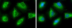
- Immunocytochemistry-Immunofluorescence analysis of ATP Citrate Lyase was performed in HeLa cells fixed in ice cold MeOH for 5 min. Green: ATP Citrate Lyase Polyclonal Antibody (Product # PA5 29497) diluted at 1:500. Blue: Hoechst 33342 staining.

- Submitted by
- Invitrogen Antibodies (provider)
- Main image
- Experimental details
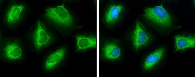
- Immunocytochemistry-Immunofluorescence analysis of ATP Citrate Lyase was performed in HeLa cells fixed in ice cold MeOH for 5 min. Green: ATP Citrate Lyase Polyclonal Antibody (Product # PA5 29497) diluted at 1:500. Blue: Hoechst 33342 staining.
Supportive validation
- Submitted by
- Invitrogen Antibodies (provider)
- Main image
- Experimental details
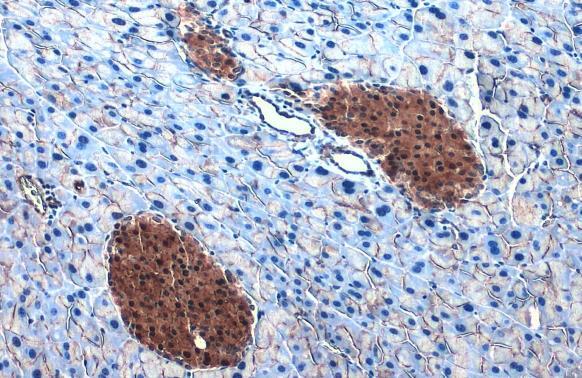
- ATP Citrate Lyase Polyclonal Antibody detects ATP citrate lyase protein at cytoplasm by immunohistochemical analysis. Sample: Paraffin-embedded mouse pancreas. ATP citrate lyase stained by ATP Citrate Lyase Polyclonal Antibody (Product # PA5-29497) diluted at 1:500. Antigen Retrieval: Citrate buffer, pH 6.0, 15 min.
- Submitted by
- Invitrogen Antibodies (provider)
- Main image
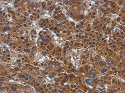
- Experimental details
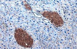
- Immunohistochemical analysis of paraffin-embedded Hepatoma, using ATP citrate lyase (Product # PA5-29497) antibody at 1:500 dilution. Antigen Retrieval: EDTA based buffer, pH 8.0, 15 min.
