Explore
 Validate
 Learn
Learn Western blot
 ELISA
ELISAAntibody data
- Antibody Data
- Antigen structure
- References [0]
- Comments [0]
- Validations
- Western blot [2]
- Other assay [1]
Submit
Validation data
Reference
Comment
Report error
- Product number
- NBP1-05215 - Provider product page

- Provider
- Novus Biologicals
- Proper citation
- Novus Cat#NBP1-05215, RRID:AB_1555290
- Product name
- Rabbit Polyclonal NF-H Antibody
- Antibody type
- Polyclonal
- Description
- Unpurified.
- Reactivity
- Rat, Chicken/Avian
- Host
- Rabbit
- Isotype
- IgG
- Vial size
- 0.1 ml
- Storage
- Store at 4C short term. Aliquot and store at -20C long term. Avoid freeze-thaw cycles.
No comments: Submit comment
Supportive validation
- Submitted by
- Novus Biologicals (provider)
- Main image

- Experimental details
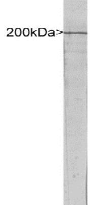
- Western Blot: NF-H Antibody [NBP1-05215] - Blot of rat spinal cord extract blotted with NBP1-05215. A major band running at 200kDa is NF-H, the major neurofilament subunit protein. A minor band at about 160kDa is the non-phosphorylated dendritic and perilaryal form of this protein.
- Submitted by
- Novus Biologicals (provider)
- Main image
- Experimental details
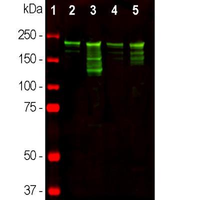
- Western Blot: NF-H Antibody [NBP1-05215] - Analysis of different tissue lysates using NF-H antibody, dilution 1:10,000 (Green): [1] protein standard (Red), [2] rat brain, [3] rat spinal cord [4] mouse brain, and [5] mouse spinal cord lysate. Strong band at about 220kDa corresponds to the phosphorylated axonal form of the NF-H subunit. Smaller proteolytic fragments of NF-H are also detected.
Supportive validation
- Submitted by
- Novus Biologicals (provider)
- Main image
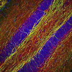
- Experimental details
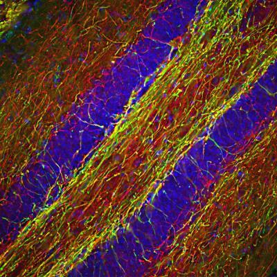
- Immunohistochemistry Free-Floating: NF-H Antibody [NBP1-05215] - Analysis of a mouse hippocampus section stained with NF-H antibody, dilution 1:2,000 (Red), and costained with mouse myelin basic protein mAb (MBP), dilution 1:5,000 (Green). DAPI staining of nuclear DNA (Blue). Following transcardial perfusion with 4% paraformaldehyde, brain was post fixed for 24hrs, cut to 45uM, and free-floating sections were stained with above antibodies. The NF-H antibody labels a network of axons of different neurons, while the MBP antibody stains myelin sheath around these axons.