 Explore
 Validate
 Learn
Learn Western blot
 Immunocytochemistry
Antibody data
- Antibody Data
- Antigen structure
- References [0]
- Comments [0]
- Validations
- Western blot [2]
- Other assay [1]
Submit
Validation data
Reference
Comment
Report error
- Product number
- NBP1-05210 - Provider product page

- Provider
- Novus Biologicals
- Proper citation
- Novus Cat#NBP1-05210, RRID:AB_1556326
- Product name
- Mouse Monoclonal NF-H Antibody
- Antibody type
- Monoclonal
- Description
- Affinity purified.
- Reactivity
- Human, Mouse, Rat
- Host
- Mouse
- Isotype
- IgG
- Vial size
- 0.1 ml
- Concentration
- 1 mg/ml
- Storage
- Store at 4C short term. Aliquot and store at -20C long term. Avoid freeze-thaw cycles.
No comments: Submit comment
Supportive validation
- Submitted by
- Novus Biologicals (provider)
- Main image
- Experimental details
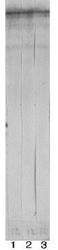
- Western Blot: NF-H Antibody (9B12) [NBP1-05210] - Strip blots of crude rat spinal cord extract stained with three different antibodies to phosphorylated NF-H, NB300-136 (lane 1), NBP1-05209 (Lane 2) and NBP1-05210 (lane 3). All three antibodies bind to a prominent band with an apparent SDS-PAGE molecular weight of 200kDa.

- Submitted by
- Novus Biologicals (provider)
- Main image
- Experimental details
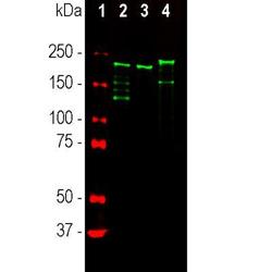
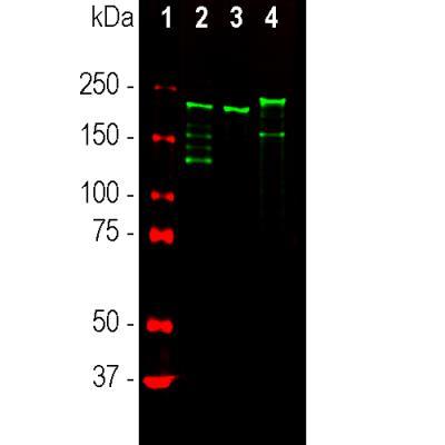
- Western Blot: NF-H Antibody (9B12) [NBP1-05210] - Analysis of different tissue lysates using mouse mAb to NF-H, dilution 1:10000 (Green): [1] protein standard, [2] rat spinal cord [3] mouse spinal cord, and [4] cow spinal cord. Strong band at about 200-220kDa corresponds to the major phosphorylated form of the NF-H subunit. Smaller proteolytic fragments of NF-H are also detected in some preparations.
Supportive validation
- Submitted by
- Novus Biologicals (provider)
- Main image
- Experimental details
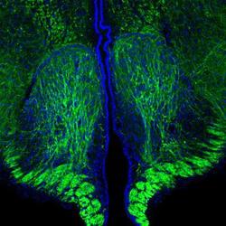
- Immunohistochemistry Free-Floating: NF-H Antibody (9B12) [NBP1-05210] - Analysis of a rat brain coronal section of the third ventricle stained with mouse monoclonal antibody to phosphorylated NF-H, dilution 1:5,000 (Green). Hoechst staining of nuclear DNA (Blue). Following transcardial perfusion with 4% paraformaldehyde, brain was post fixed for 24hrs, cut to 45uM, and free-floating sections were stained with above antibody. The antibody is a robust marker of the axons of neuronal cells.
