 Explore
 Validate
 Learn
Learn Western blot
Antibody data
- Antibody Data
- Antigen structure
- References [2]
- Comments [0]
- Validations
- Western blot [1]
- Immunohistochemistry [1]
Submit
Validation data
Reference
Comment
Report error
- Product number
- MA1-10041 - Provider product page

- Provider
- Invitrogen Antibodies
- Product name
- NF-H Monoclonal Antibody (NAP4)
- Antibody type
- Monoclonal
- Antigen
- Purifed from natural sources
- Description
- MA1-10041 recognizes phosphorylated NF-H KSP (lysine-serine-proline) type sequences.
- Reactivity
- Human, Mouse, Rat, Bovine, Chicken/Avian, Porcine
- Host
- Mouse
- Isotype
- IgG
- Antibody clone number
- NAP4
- Vial size
- 100 µL
- Concentration
- 1 mg/mL
- Storage
- Store at 4°C short term. For long term storage, store at -20°C, avoiding freeze/thaw cycles.
Submitted references Compartmentation of alpha-internexin and neurofilament triplet proteins in cultured hippocampal neurons.
Compartmentation of alpha-internexin and neurofilament triplet proteins in cultured hippocampal neurons.
Benson DL, Mandell JW, Shaw G, Banker G
Journal of neurocytology 1996 Mar;25(3):181-96
Journal of neurocytology 1996 Mar;25(3):181-96
Compartmentation of alpha-internexin and neurofilament triplet proteins in cultured hippocampal neurons.
Benson DL, Mandell JW, Shaw G, Banker G
Journal of neurocytology 1996 Mar;25(3):181-96
Journal of neurocytology 1996 Mar;25(3):181-96
No comments: Submit comment
Supportive validation
- Submitted by
- Invitrogen Antibodies (provider)
- Main image
- Experimental details
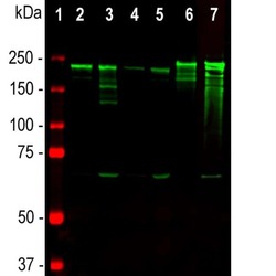
- Western blot analysis of NF-H in tissue lysates using an NF-H monoclonal antibody (Product # MA1-10041) at a dilution of 1:10,000 seen in green. 1) protein standard (red), 2) rat brain, 3) rat spinal cord, 4) mouse brain, 5) mouse spinal cord, 6) pig spinal cord, 7) cow spinal cord. Strong band at about 200-220 kDa corresponds to the major phosphorylated form of the NF-H subunit. A minor band at about 160 kDa is the non-phosphorylated NF-H. Smaller proteolytic fragments of NF-H are also detected in spinal cord preparations with the antibody.

Supportive validation
- Submitted by
- Invitrogen Antibodies (provider)
- Main image

- Experimental details
- Immunohistological analysis of NF-H in human cerebellar cortex section. Paraffin-embedded, formalin-fixed tissue sections were stained with an NF-H monoclonal antibody (Product # MA1-10041) seen in brown and counterstained with Hematoxylin in blue. The antibody stains prominent basket cell axons surrounding the large Purkinje neurons.
