Explore
 Validate
 Learn
Learn Western blot
Antibody data
- Antibody Data
- Antigen structure
- References [1]
- Comments [0]
- Validations
- Western blot [3]
- Immunocytochemistry [1]
- Immunohistochemistry [5]
Submit
Validation data
Reference
Comment
Report error
- Product number
- GTX110065 - Provider product page

- Provider
- GeneTex
- Proper citation
- GeneTex Cat#GTX110065, RRID:AB_1950985
- Product name
- NF-H antibody
- Antibody type
- Polyclonal
- Reactivity
- Human, Mouse, Rat
- Host
- Rabbit
Submitted references Histone deacetylase 4 protects from denervation and skeletal muscle atrophy in a murine model of amyotrophic lateral sclerosis.
Pigna E, Simonazzi E, Sanna K, Bernadzki KM, Proszynski T, Heil C, Palacios D, Adamo S, Moresi V
EBioMedicine 2019 Feb;40:717-732
EBioMedicine 2019 Feb;40:717-732
No comments: Submit comment
Supportive validation
- Submitted by
- GeneTex (provider)
- Main image
- Experimental details
- Sample (30 ug of whole cell lysate) A: Hela 7.5% SDS PAGE GTX110065 diluted at 1:1000
- Validation comment
- WB
- Submitted by
- GeneTex (provider)
- Main image
- Experimental details
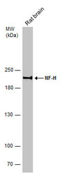
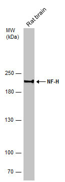
- Rat tissue extract (50 ?g) was separated by 5% SDS-PAGE, and the membrane was blotted with NF-H antibody (GTX110065) diluted at 1:500.
- Submitted by
- GeneTex (provider)
- Main image
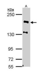
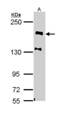
- Experimental details
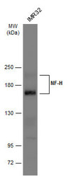
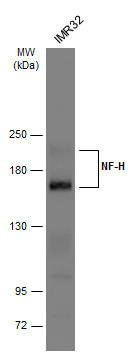
- Whole cell extract (30 ?g) was separated by 5% SDS-PAGE, and the membrane was blotted with NF-H antibody (GTX110065) diluted at 1:500. The HRP-conjugated anti-rabbit IgG antibody (GTX213110-01) was used to detect the primary antibody.
Supportive validation
- Submitted by
- GeneTex (provider)
- Main image
- Experimental details
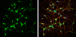
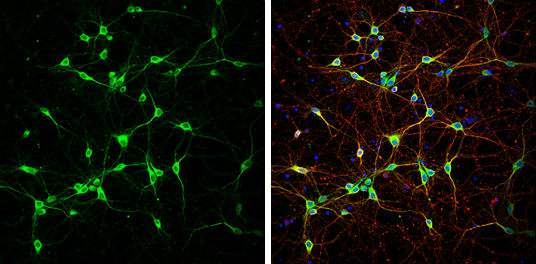
- NF-H antibody detects NF-H protein at cytoplasm by immunofluorescent analysis.Sample: DIV9 rat E18 primary cortical neurons were fixed in 4% paraformaldehyde at RT for 15 min.Green: NF-H protein stained by NF-H antibody (GTX110065) diluted at 1:500.Red: beta Tubulin 3/ Tuj1, stained by beta Tubulin 3/ Tuj1 antibody [GT11710] (GTX631836) diluted at 1:500.Blue: Fluoroshield with DAPI (GTX30920).
Supportive validation
- Submitted by
- GeneTex (provider)
- Main image

- Experimental details
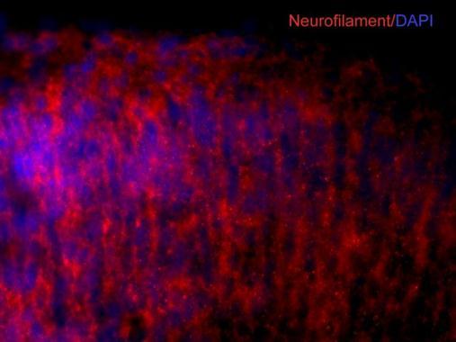
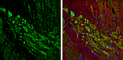
- NF-H antibody detects NF-H proteins in embryonic mouse brain by immunohistochemical analysis. Sample: Frozen section of embryonic mouse brain (mE18.5). Red: NF-H antibody (GTX110065) diluted at 1:500. Blue: DAPI.
- Submitted by
- GeneTex (provider)
- Main image

- Experimental details
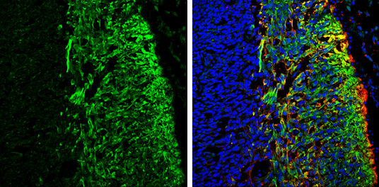
- NF-H antibody detects NF-H protein expression by immunohistochemical analysis.Sample: Frozen sectioned E13.5 Rat brain. Green: NF-H protein stained by NF-H antibody (GTX110065) diluted at 1:250.Red: beta Tubulin 3/ TUJ1, a mature neuron marker, stained by beta Tubulin 3/ TUJ1 antibody [GT11710] (GTX631836) diluted at 1:500.Blue: Fluoroshield with DAPI (GTX30920).
- Submitted by
- GeneTex (provider)
- Main image
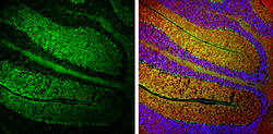
- Experimental details
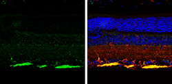
- NF-H antibody detects NF-H protein expression by immunohistochemical analysis.Sample:Paraffin-Embedded adult mouse retina. Green: NF-H protein stained by NF-H antibody (GTX110065) diluted at 1:250.Red: beta Tubulin 3/ TUJ1, stained by beta Tubulin 3/ TUJ1 antibody [GT11710] (GTX631836) diluted at 1:500.Blue: Fluoroshield with DAPI (GTX30920).

- Submitted by
- GeneTex (provider)
- Main image
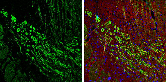
- Experimental details
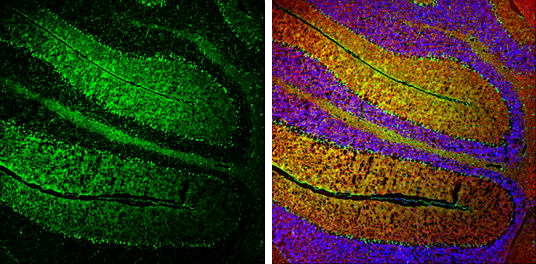
- NF-H antibody detects NF-H protein expression by immunohistochemical analysis.Sample: Frozen-sectioned adult mouse cerebellum. Green: NF-H protein stained by NF-H antibody (GTX110065) diluted at 1:250.Red: beta Tubulin 3/ TUJ1, stained by beta Tubulin 3/ TUJ1 antibody [GT11710] (GTX631836) diluted at 1:500.Blue: Fluoroshield with DAPI (GTX30920).
- Submitted by
- GeneTex (provider)
- Main image
- Experimental details
- NF-H antibody detects NF-H protein expression by immunohistochemical analysis.Sample: Frozen-sectioned adult mouse cerebellum. Green: NF-H protein stained by NF-H antibody (GTX110065) diluted at 1:250.Red: beta Tubulin 3/ TUJ1, stained by beta Tubulin 3/ TUJ1 antibody [GT11710] (GTX631836) diluted at 1:500.Blue: Fluoroshield with DAPI (GTX30920).