Explore
 Validate
 Learn
Learn Western blot
 Immunocytochemistry
Antibody data
- Antibody Data
- Antigen structure
- References [0]
- Comments [0]
- Validations
- Immunocytochemistry [4]
Submit
Validation data
Reference
Comment
Report error
- Product number
- LS-C204550 - Provider product page

- Provider
- LSBio
- Product name
- NEFH / NF-H Antibody (clone AH1) LS-C204550
- Antibody type
- Monoclonal
- Description
- Ascites
- Reactivity
- Bovine, Chicken/Avian
- Host
- Mouse
- Isotype
- IgG
- Antibody clone number
- AH1
- Storage
- Store at 4°C or -20°C. Avoid freeze-thaw cycles.
No comments: Submit comment
Enhanced validation
- Submitted by
- LSBio (provider)
- Enhanced method
- Genetic validation
- Main image

- Experimental details
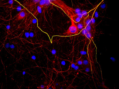
- Mixed neuron/glial cultures stained with NEFH / NF-H antibody (green) and also stained with rabbit polyclonal antibody to neurofilament NF-L RPCA-NF-L (red). The NF-L antibody stains neurofilaments in both axons and dendrites, and so can reveal neuronal cell bodies, while NEFH / NF-H antibody binds to only heavily phosphorylated forms of NF-H which are localized to mature axonal neurofilaments. In this image a few axons course from left to right and top to bottom- since they contain both NF-L and phosphorylated NF-H they appear golden in color. Blue shows the distribution of DNA.
- Submitted by
- LSBio (provider)
- Enhanced method
- Genetic validation
- Main image

- Experimental details
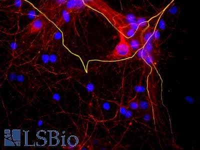
- Mixed neuron/glial cultures stained with NEFH / NF-H antibody (green) and also stained with rabbit polyclonal antibody to neurofilament NF-L RPCA-NF-L (red). The NF-L antibody stains neurofilaments in both axons and dendrites, and so can reveal neuronal cell bodies, while NEFH / NF-H antibody binds to only heavily phosphorylated forms of NF-H which are localized to mature axonal neurofilaments. In this image a few axons course from left to right and top to bottom- since they contain both NF-L and phosphorylated NF-H they appear golden in color. Blue shows the distribution of DNA.

- Submitted by
- LSBio (provider)
- Main image

- Experimental details
- Mixed neuron/glial cultures stained with NEFH / NF-H antibody (green) and also stained with rabbit polyclonal antibody to neurofilament NF-L RPCA-NF-L (red). The NF-L antibody stains neurofilaments in both axons and dendrites, and so can reveal neuronal cell bodies, while NEFH / NF-H antibody binds to only heavily phosphorylated forms of NF-H which are localized to mature axonal neurofilaments. In this image a few axons course from left to right and top to bottom- since they contain both NF-L and phosphorylated NF-H they appear golden in color. Blue shows the distribution of DNA.