 Explore
 Validate
 Learn
Learn Western blot
 Immunocytochemistry
 Immunohistochemistry
Antibody data
- Antibody Data
- Antigen structure
- References [0]
- Comments [0]
- Validations
- Western blot [1]
- Immunohistochemistry [2]
Submit
Validation data
Reference
Comment
Report error
- Product number
- LS-C204551 - Provider product page

- Provider
- LSBio
- Product name
- NEFH / NF-H Antibody (clone NAP4) LS-C204551
- Antibody type
- Monoclonal
- Description
- Ascites
- Reactivity
- Chicken/Avian, Porcine
- Host
- Mouse
- Isotype
- IgG
- Antibody clone number
- NAP4
- Storage
- Store at 4°C or -20°C. Avoid freeze-thaw cycles.
No comments: Submit comment
Enhanced validation
- Submitted by
- LSBio (provider)
- Enhanced method
- Genetic validation
- Main image

- Experimental details
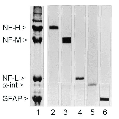
- Rat spinal cord homogenate showing the major intermediate filament proteins of the nervous system (lane 1). The remaining lanes show blots of this material stained with various antibodies including NEFH / NF-H antibody (lane 2).
Supportive validation
- Submitted by
- LSBio (provider)
- Enhanced method
- Genetic validation
- Main image
- Experimental details
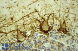
- Human cerebellar cortex fixed in formalin, embedded in paraffin and stained with NEFH / NF-H antibody using the ABC (avidin biotin conjugate) method. The section was counterstained with hematoxylin-Eosin (blue). NEFH / NF-H antibody stains prominent basket cell axons surrounding the large Purkinje neurons. Granule cell layer at bottom of image, molecular layer at top.

- Submitted by
- LSBio (provider)
- Enhanced method
- Genetic validation
- Main image

- Experimental details
- Human cerebellar cortex fixed in formalin, embedded in paraffin and stained with NEFH / NF-H antibody using the ABC (avidin biotin conjugate) method. The section was counterstained with hematoxylin-Eosin (blue). NEFH / NF-H antibody stains prominent basket cell axons surrounding the large Purkinje neurons. Granule cell layer at bottom of image, molecular layer at top.
