 Explore
 Validate
 Learn
Learn Western blot
 Immunocytochemistry
Antibody data
- Antibody Data
- Antigen structure
- References [1]
- Comments [0]
- Validations
- Western blot [1]
- Immunohistochemistry [1]
Submit
Validation data
Reference
Comment
Report error
- Product number
- NB300-136 - Provider product page

- Provider
- Novus Biologicals
- Proper citation
- Novus Cat#NB300-136, RRID:AB_10002635
- Product name
- Mouse Monoclonal NF-H Antibody
- Antibody type
- Monoclonal
- Description
- Affinity purified. Specifically recognizes the phosphorylated variant of NF-H subunit (~200-220 kDa), showing some weaker reactivity with phosphorylated forms of NF-M.
- Reactivity
- Human, Mouse, Rat, Bovine, Chicken/Avian, Porcine
- Host
- Mouse
- Isotype
- IgG
- Vial size
- 0.1 ml
- Concentration
- 1 mg/ml
- Storage
- Store at 4C short term. Aliquot and store at -20C long term. Avoid freeze-thaw cycles.
Submitted references Caveolin-1 deficiency increases cerebral ischemic injury.
Jasmin JF, Malhotra S, Singh Dhallu M, Mercier I, Rosenbaum DM, Lisanti MP
Circulation research 2007 Mar 16;100(5):721-9
Circulation research 2007 Mar 16;100(5):721-9
No comments: Submit comment
Supportive validation
- Submitted by
- Novus Biologicals (provider)
- Main image
- Experimental details
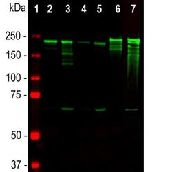
- Western Blot: NF-H Antibody (NAP4) [NB300-136] - Analysis of tissue lysates using mouse mAb to NF-H, NB300-136, dilution 1:10,000 in green: [1] protein standard (red), [2] rat brain, [3] rat spinal cord, [4] mouse brain, [5] mouse spinal cord, [6] pig spinal cord, [7] cow spinal cord. Strong band at about 200-220 kDa corresponds to the major phosphorylated from of the NF-H subunit. A minor band at about 160 kDa is the non-phosphorylated NF-H. Smaller proteolytic fragments of NF-H are also detected in spinal cord preparations with NB300-136 antibody.

Supportive validation
- Submitted by
- Novus Biologicals (provider)
- Main image

- Experimental details
- Immunohistochemistry: NF-H Antibody (NAP4) [NB300-136] - Analysis of human cerebellar cortex section stained with mouse mAb to pNF-H, NB300-136, in brown. Paraffin-embedded, formalin-fixed tissue sections were stained with this antibody using the avidin biotin conjugate method. The sections was counterstained with Hematoxylin in blue. NB300-136 stains prominent basket cell axons surrounding the large Purkinje neurons. Cerebellar granule cell layer is at the bottom of the image, the molecular layer at the top.
