 Explore
 Validate
 Learn
Learn Western blot
 ELISA
ELISAAntibody data
- Antibody Data
- Antigen structure
- References [3]
- Comments [0]
- Validations
- Western blot [2]
- Immunohistochemistry [1]
- Other assay [1]
Submit
Validation data
Reference
Comment
Report error
- Product number
- NB300-217 - Provider product page

- Provider
- Novus Biologicals
- Proper citation
- Novus Cat#NB300-217, RRID:AB_2149775
- Product name
- Chicken Polyclonal NF-H Antibody
- Antibody type
- Polyclonal
- Description
- Ammonium sulfate precipitation. Reacts very strongly with NF-H KSP type phosphorylated repeats. Reactivity with non-phosphorylated KSP sequences is orders of magnitude weaker.
- Reactivity
- Human, Mouse, Rat, Bovine, Canine, Feline, Porcine
- Host
- Chicken/Avian
- Isotype
- IgY
- Vial size
- 0.05 ml
- Storage
- Store at 4C short term. Aliquot and store at -20C long term. Avoid freeze-thaw cycles.
Submitted references Interactions between mitochondria and endoplasmic reticulum in demyelinated axons.
Controlling the dose-dependent, synergistic and temporal effects of NGF and GDNF by encapsulation in PLGA microparticles for use in nerve guidance conduits for the repair of large peripheral nerve defects.
Afferent Innervation, Muscle Spindles, and Contractures Following Neonatal Brachial Plexus Injury in a Mouse Model.
Thai TQ, Nguyen HB, Sui Y, Ikenaka K, Oda T, Ohno N
Medical molecular morphology 2019 Sep;52(3):135-146
Medical molecular morphology 2019 Sep;52(3):135-146
Controlling the dose-dependent, synergistic and temporal effects of NGF and GDNF by encapsulation in PLGA microparticles for use in nerve guidance conduits for the repair of large peripheral nerve defects.
Lackington WA, Kočí Z, Alekseeva T, Hibbitts AJ, Kneafsey SL, Chen G, O'Brien FJ
Journal of controlled release : official journal of the Controlled Release Society 2019 Jun 28;304:51-64
Journal of controlled release : official journal of the Controlled Release Society 2019 Jun 28;304:51-64
Afferent Innervation, Muscle Spindles, and Contractures Following Neonatal Brachial Plexus Injury in a Mouse Model.
Nikolaou S, Hu L, Cornwall R
The Journal of hand surgery 2015 Oct;40(10):2007-16
The Journal of hand surgery 2015 Oct;40(10):2007-16
No comments: Submit comment
Supportive validation
- Submitted by
- Novus Biologicals (provider)
- Main image

- Experimental details
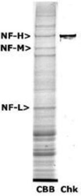
- Western Blot: NF-H Antibody [NB300-217] - Analysis of 200kDa Neurofilament Heavy expression in rat spinal cord extract. The first lane is Coomassie Brilliant Blue stained and the second lane is probed with chicken anti-Neurofilament Heavy antibody NB300-217.
- Submitted by
- Novus Biologicals (provider)
- Main image
- Experimental details
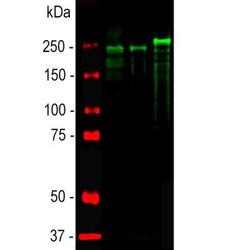
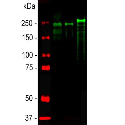
- Western Blot: NF-H Antibody [NB300-217] - Analysis of spinal cord lysates from different species using chicken pAb to NF-H, NB300-217, dilution 1:20,000 in green: [1] protein standard (red), [2] rat, [3] mouse, and [4] cow spinal cord. Strong band at about 200-220kDa corresponds to the phosphorylated from of NF-H. The protein from different species is known to have different SDS-PAGE molecular weights, with large species generally expressing larger proteins. Smaller proteolytic fragments of NF-H are also detected in spinal cord preparations with this antibody. The antibody does not recognize non-phosphorylated forms of NF-H.
Supportive validation
- Submitted by
- Novus Biologicals (provider)
- Main image
- Experimental details
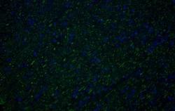
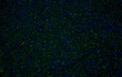
- Immunohistochemistry-Frozen: NF-H Antibody [NB300-217] - Canine optic nerve, antibody specifically labelled the axons (Green). Blue is DAPI. Image taken with an epifluorescent microscope and was incubated at 1:10000 for 1hr at RT.. Image from verified customer review.
Supportive validation
- Submitted by
- Novus Biologicals (provider)
- Main image
- Experimental details
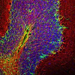
- Immunohistochemistry Free-Floating: NF-H Antibody [NB300-217] - Analysis of a rat cerebellum section stained with NF-H antibody, dilution 1:5,000 (Red), and costained with rabbit GFAP pAb, dilution 1:5,000 Green). DAPI staining of nuclear DNA (Blue). Following transcardial perfusion with 4% paraformaldehyde, brain was post fixed for 24hrs, cut to 45uM, and free floating sections were stained with above antibodies. The NF-H antibody labels network of axons of different neurons, while the GFAP antibody stains astrocytes and other glial cells.
