 Explore
 Validate
 Learn
Learn Western blot
Antibody data
- Antibody Data
- Antigen structure
- References [0]
- Comments [0]
- Validations
- Western blot [1]
- Immunohistochemistry [39]
Submit
Validation data
Reference
Comment
Report error
- Product number
- LS-C483021 - Provider product page

- Provider
- LSBio
- Product name
- NEFH / NF-H Antibody (aa150-200) LS-C483021
- Antibody type
- Polyclonal
- Description
- Antiserum
- Reactivity
- Human, Mouse, Rat
- Host
- Rabbit
- Storage
- Maintain lyophilized and reconstituted antibodies at -20°C for long term storage and at 2°C to 8°C for a shorter term. When reconstituting, glycerol (1:1) may be added for an additional stability. Avoid freeze/thaw cycles.
No comments: Submit comment
Enhanced validation
- Submitted by
- LSBio (provider)
- Enhanced method
- Genetic validation
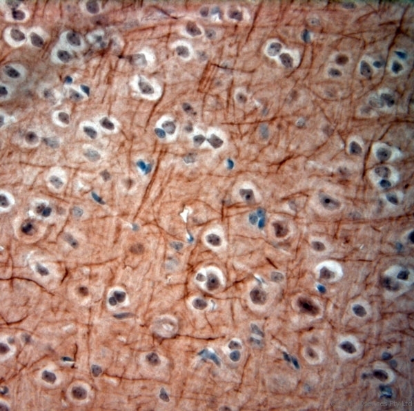
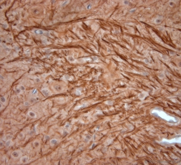
- Main image

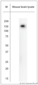
- Experimental details
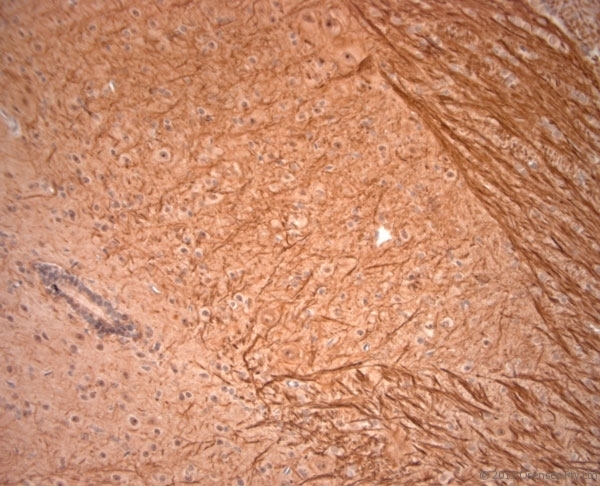
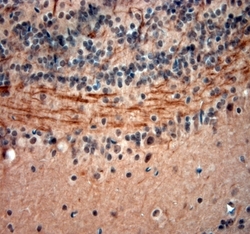
- Rabbit antibody to 200 Neurofilament (150-200). WB on mouse brain lysate. Blocking: 1% LFDM for 30 min at RT; primary antibody: dilution 1:1000 incubated at 4C overnight.
Supportive validation
- Submitted by
- LSBio (provider)
- Enhanced method
- Genetic validation
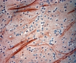
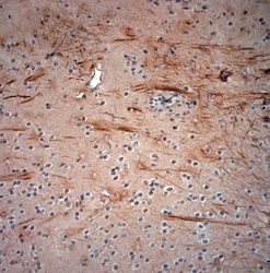
- Main image

- Experimental details
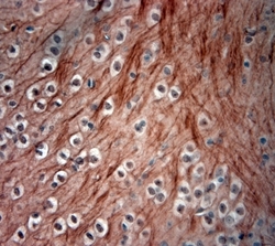
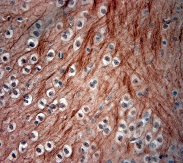
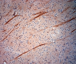
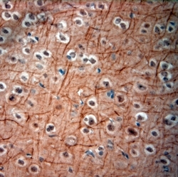
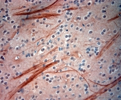
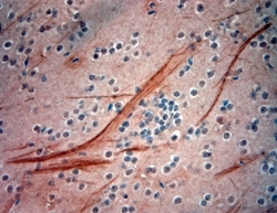
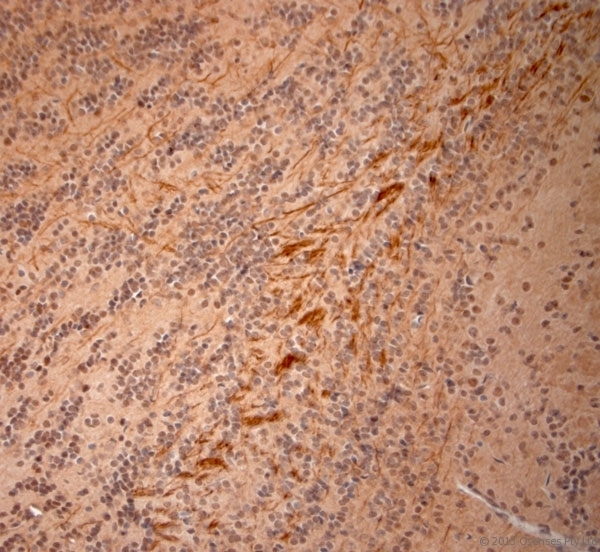
- Rabbit antibody to 200 Neurofilament (150-200). IHC-P on paraffin sections of mouse spinal cord. The animal was perfused using Autoperfuser at a pressure of 110 mm Hg with 300 ml 4% FA and further post fixed overnight before being processed for paraffin embedding. HIER: Tris-EDTA, pH 9 for 20 min using Thermo PT Module. Blocking: 0.2% LFDM in TBST filtered through a 0.2 micron filter. Detection was done using Novolink HRP polymer from Leica following manufacturers instructions, using DAB chromogen. Primary antibody: dilution 1:1000, incubated 30 min at RT using Autostainer. Sections were counterstained with Harris Hematoxylin.

- Submitted by
- LSBio (provider)
- Enhanced method
- Genetic validation
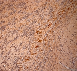
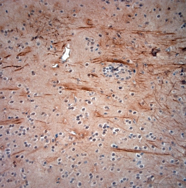
- Main image

- Experimental details
- Rabbit antibody to 200 Neurofilament (150-200). IHC-P on paraffin sections of mouse spinal cord. The animal was perfused using Autoperfuser at a pressure of 110 mm Hg with 300 ml 4% FA and further post fixed overnight before being processed for paraffin embedding. HIER: Tris-EDTA, pH 9 for 20 min using Thermo PT Module. Blocking: 0.2% LFDM in TBST filtered through a 0.2 micron filter. Detection was done using Novolink HRP polymer from Leica following manufacturers instructions, using DAB chromogen. Primary antibody: dilution 1:1000, incubated 30 min at RT using Autostainer. Sections were counterstained with Harris Hematoxylin.

- Submitted by
- LSBio (provider)
- Enhanced method
- Genetic validation
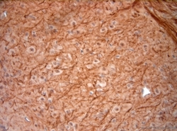
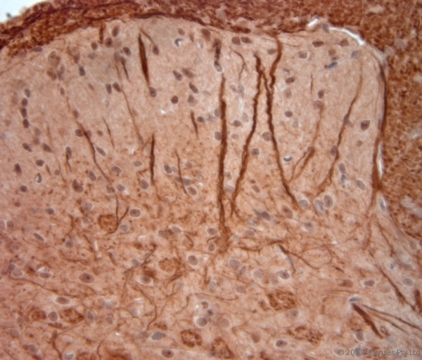
- Main image
- Experimental details
- Rabbit antibody to 200 Neurofilament (150-200). IHC-P on paraffin sections of mouse spinal cord. The animal was perfused using Autoperfuser at a pressure of 110 mm Hg with 300 ml 4% FA and further post fixed overnight before being processed for paraffin embedding. HIER: Tris-EDTA, pH 9 for 20 min using Thermo PT Module. Blocking: 0.2% LFDM in TBST filtered through a 0.2 micron filter. Detection was done using Novolink HRP polymer from Leica following manufacturers instructions, using DAB chromogen. Primary antibody: dilution 1:1000, incubated 30 min at RT using Autostainer. Sections were counterstained with Harris Hematoxylin.

- Submitted by
- LSBio (provider)
- Enhanced method
- Genetic validation
- Main image

- Experimental details
- Rabbit antibody to 200 Neurofilament (150-200). IHC-P on paraffin sections of mouse spinal cord. The animal was perfused using Autoperfuser at a pressure of 110 mm Hg with 300 ml 4% FA and further post fixed overnight before being processed for paraffin embedding. HIER: Tris-EDTA, pH 9 for 20 min using Thermo PT Module. Blocking: 0.2% LFDM in TBST filtered through a 0.2 micron filter. Detection was done using Novolink HRP polymer from Leica following manufacturers instructions, using DAB chromogen. Primary antibody: dilution 1:1000, incubated 30 min at RT using Autostainer. Sections were counterstained with Harris Hematoxylin.
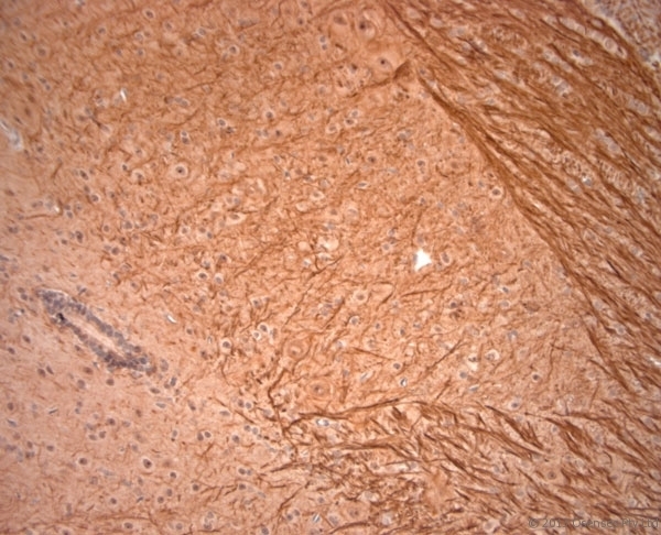
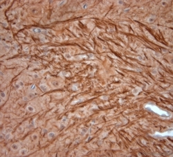
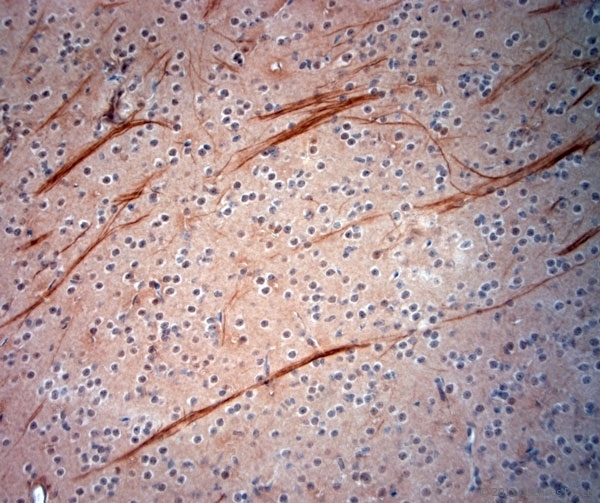
- Submitted by
- LSBio (provider)
- Enhanced method
- Genetic validation
- Main image

- Experimental details
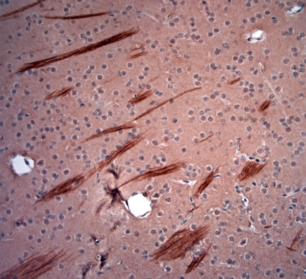
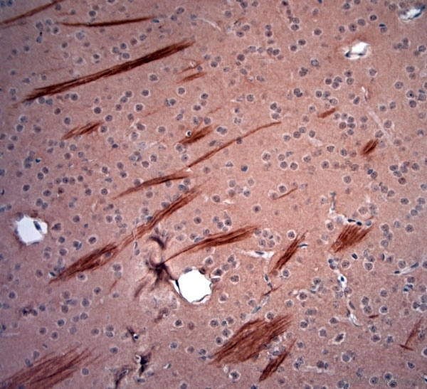
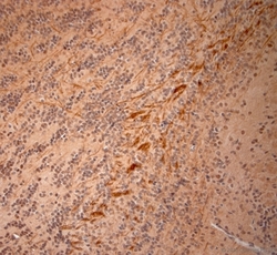
- Rabbit antibody to 200 Neurofilament (150-200). IHC-P on paraffin sections of mouse brain. The animal was perfused using Autoperfuser at a pressure of 110 mm Hg with 300 ml 4% FA and further post fixed overnight before being processed for paraffin embedding. HIER: Tris-EDTA, pH 9 for 20 min using Thermo PT Module. Blocking: 0.2% LFDM in TBST filtered through a 0.2 micron filter. Detection was done using Novolink HRP polymer from Leica following manufacturers instructions, using DAB chromogen. Primary antibody: dilution 1:1000, incubated 30 min at RT using Autostainer. Sections were counterstained with Harris Hematoxylin.

- Submitted by
- LSBio (provider)
- Enhanced method
- Genetic validation
- Main image

- Experimental details
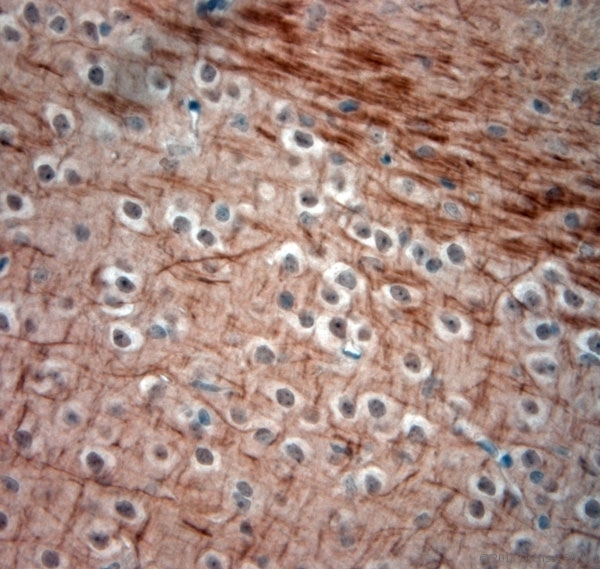
- Rabbit antibody to 200 Neurofilament (150-200). IHC-P on paraffin sections of mouse brain. The animal was perfused using Autoperfuser at a pressure of 110 mm Hg with 300 ml 4% FA and further post fixed overnight before being processed for paraffin embedding. HIER: Tris-EDTA, pH 9 for 20 min using Thermo PT Module. Blocking: 0.2% LFDM in TBST filtered through a 0.2 micron filter. Detection was done using Novolink HRP polymer from Leica following manufacturers instructions, using DAB chromogen. Primary antibody: dilution 1:1000, incubated 30 min at RT using Autostainer. Sections were counterstained with Harris Hematoxylin.
- Submitted by
- LSBio (provider)
- Enhanced method
- Genetic validation
- Main image

- Experimental details
- Rabbit antibody to 200 Neurofilament (150-200). IHC-P on paraffin sections of mouse brain. The animal was perfused using Autoperfuser at a pressure of 110 mm Hg with 300 ml 4% FA and further post fixed overnight before being processed for paraffin embedding. HIER: Tris-EDTA, pH 9 for 20 min using Thermo PT Module. Blocking: 0.2% LFDM in TBST filtered through a 0.2 micron filter. Detection was done using Novolink HRP polymer from Leica following manufacturers instructions, using DAB chromogen. Primary antibody: dilution 1:1000, incubated 30 min at RT using Autostainer. Sections were counterstained with Harris Hematoxylin.
- Submitted by
- LSBio (provider)
- Enhanced method
- Genetic validation
- Main image
- Experimental details
- Rabbit antibody to 200 Neurofilament (150-200). IHC-P on paraffin sections of mouse brain. The animal was perfused using Autoperfuser at a pressure of 110 mm Hg with 300 ml 4% FA and further post fixed overnight before being processed for paraffin embedding. HIER: Tris-EDTA, pH 9 for 20 min using Thermo PT Module. Blocking: 0.2% LFDM in TBST filtered through a 0.2 micron filter. Detection was done using Novolink HRP polymer from Leica following manufacturers instructions, using DAB chromogen. Primary antibody: dilution 1:1000, incubated 30 min at RT using Autostainer. Sections were counterstained with Harris Hematoxylin.
- Submitted by
- LSBio (provider)
- Enhanced method
- Genetic validation
- Main image

- Experimental details
- Rabbit antibody to 200 Neurofilament (150-200). IHC-P on paraffin sections of mouse brain. The animal was perfused using Autoperfuser at a pressure of 110 mm Hg with 300 ml 4% FA and further post fixed overnight before being processed for paraffin embedding. HIER: Tris-EDTA, pH 9 for 20 min using Thermo PT Module. Blocking: 0.2% LFDM in TBST filtered through a 0.2 micron filter. Detection was done using Novolink HRP polymer from Leica following manufacturers instructions, using DAB chromogen. Primary antibody: dilution 1:1000, incubated 30 min at RT using Autostainer. Sections were counterstained with Harris Hematoxylin.
- Submitted by
- LSBio (provider)
- Enhanced method
- Genetic validation
- Main image
- Experimental details
- Rabbit antibody to 200 Neurofilament (150-200). IHC-P on paraffin sections of mouse brain. The animal was perfused using Autoperfuser at a pressure of 110 mm Hg with 300 ml 4% FA and further post fixed overnight before being processed for paraffin embedding. HIER: Tris-EDTA, pH 9 for 20 min using Thermo PT Module. Blocking: 0.2% LFDM in TBST filtered through a 0.2 micron filter. Detection was done using Novolink HRP polymer from Leica following manufacturers instructions, using DAB chromogen. Primary antibody: dilution 1:1000, incubated 30 min at RT using Autostainer. Sections were counterstained with Harris Hematoxylin.

- Submitted by
- LSBio (provider)
- Enhanced method
- Genetic validation
- Main image
- Experimental details
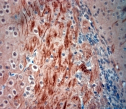
- Rabbit antibody to 200 Neurofilament (150-200). IHC-P on paraffin sections of mouse olfactory bulb. The animal was perfused using Autoperfuser at a pressure of 110 mm Hg with 300 ml 4% FA and further post fixed overnight before being processed for paraffin embedding. HIER: Tris-EDTA, pH 9 for 20 min using Thermo PT Module. Blocking: 0.2% LFDM in TBST filtered through a 0.2 micron filter. Detection was done using Novolink HRP polymer from Leica following manufacturers instructions, using DAB chromogen. Primary antibody: dilution 1:1000, incubated 30 min at RT using Autostainer. Sections were counterstained with Harris Hematoxylin.

- Submitted by
- LSBio (provider)
- Enhanced method
- Genetic validation
- Main image

- Experimental details
- Rabbit antibody to 200 Neurofilament (150-200). IHC-P on paraffin sections of mouse olfactory bulb. The animal was perfused using Autoperfuser at a pressure of 110 mm Hg with 300 ml 4% FA and further post fixed overnight before being processed for paraffin embedding. HIER: Tris-EDTA, pH 9 for 20 min using Thermo PT Module. Blocking: 0.2% LFDM in TBST filtered through a 0.2 micron filter. Detection was done using Novolink HRP polymer from Leica following manufacturers instructions, using DAB chromogen. Primary antibody: dilution 1:1000, incubated 30 min at RT using Autostainer. Sections were counterstained with Harris Hematoxylin.

- Submitted by
- LSBio (provider)
- Enhanced method
- Genetic validation
- Main image
- Experimental details
- Rabbit antibody to 200 Neurofilament (150-200). IHC-P on paraffin sections of mouse spinal cord. The animal was perfused using Autoperfuser at a pressure of 110 mm Hg with 300 ml 4% FA and further post fixed overnight before being processed for paraffin embedding. HIER: Tris-EDTA, pH 9 for 20 min using Thermo PT Module. Blocking: 0.2% LFDM in TBST filtered through a 0.2 micron filter. Detection was done using Novolink HRP polymer from Leica following manufacturers instructions, using DAB chromogen. Primary antibody: dilution 1:1000, incubated 30 min at RT using Autostainer. Sections were counterstained with Harris Hematoxylin.

- Submitted by
- LSBio (provider)
- Enhanced method
- Genetic validation
- Main image

- Experimental details
- Rabbit antibody to 200 Neurofilament (150-200). IHC-P on paraffin sections of mouse spinal cord. The animal was perfused using Autoperfuser at a pressure of 110 mm Hg with 300 ml 4% FA and further post fixed overnight before being processed for paraffin embedding. HIER: Tris-EDTA, pH 9 for 20 min using Thermo PT Module. Blocking: 0.2% LFDM in TBST filtered through a 0.2 micron filter. Detection was done using Novolink HRP polymer from Leica following manufacturers instructions, using DAB chromogen. Primary antibody: dilution 1:1000, incubated 30 min at RT using Autostainer. Sections were counterstained with Harris Hematoxylin.

- Submitted by
- LSBio (provider)
- Enhanced method
- Genetic validation
- Main image
- Experimental details
- Rabbit antibody to 200 Neurofilament (150-200). IHC-P on paraffin sections of mouse brain. The animal was perfused using Autoperfuser at a pressure of 110 mm Hg with 300 ml 4% FA and further post fixed overnight before being processed for paraffin embedding. HIER: Tris-EDTA, pH 9 for 20 min using Thermo PT Module. Blocking: 0.2% LFDM in TBST filtered through a 0.2 micron filter. Detection was done using Novolink HRP polymer from Leica following manufacturers instructions, using DAB chromogen. Primary antibody: dilution 1:1000, incubated 30 min at RT using Autostainer. Sections were counterstained with Harris Hematoxylin.
- Submitted by
- LSBio (provider)
- Enhanced method
- Genetic validation
- Main image

- Experimental details
- Rabbit antibody to 200 Neurofilament (150-200). IHC-P on paraffin sections of mouse brain. The animal was perfused using Autoperfuser at a pressure of 110 mm Hg with 300 ml 4% FA and further post fixed overnight before being processed for paraffin embedding. HIER: Tris-EDTA, pH 9 for 20 min using Thermo PT Module. Blocking: 0.2% LFDM in TBST filtered through a 0.2 micron filter. Detection was done using Novolink HRP polymer from Leica following manufacturers instructions, using DAB chromogen. Primary antibody: dilution 1:1000, incubated 30 min at RT using Autostainer. Sections were counterstained with Harris Hematoxylin.

- Submitted by
- LSBio (provider)
- Enhanced method
- Genetic validation
- Main image

- Experimental details
- Rabbit antibody to 200 Neurofilament (150-200). IHC-P on paraffin sections of mouse olfactory bulb. The animal was perfused using Autoperfuser at a pressure of 110 mm Hg with 300 ml 4% FA and further post fixed overnight before being processed for paraffin embedding. HIER: Tris-EDTA, pH 9 for 20 min using Thermo PT Module. Blocking: 0.2% LFDM in TBST filtered through a 0.2 micron filter. Detection was done using Novolink HRP polymer from Leica following manufacturers instructions, using DAB chromogen. Primary antibody: dilution 1:1000, incubated 30 min at RT using Autostainer. Sections were counterstained with Harris Hematoxylin.

- Submitted by
- LSBio (provider)
- Enhanced method
- Genetic validation
- Main image

- Experimental details
- Rabbit antibody to 200 Neurofilament (150-200). IHC-P on paraffin sections of mouse spinal cord. The animal was perfused using Autoperfuser at a pressure of 110 mm Hg with 300 ml 4% FA and further post fixed overnight before being processed for paraffin embedding. HIER: Tris-EDTA, pH 9 for 20 min using Thermo PT Module. Blocking: 0.2% LFDM in TBST filtered through a 0.2 micron filter. Detection was done using Novolink HRP polymer from Leica following manufacturers instructions, using DAB chromogen. Primary antibody: dilution 1:1000, incubated 30 min at RT using Autostainer. Sections were counterstained with Harris Hematoxylin.
- Submitted by
- LSBio (provider)
- Enhanced method
- Genetic validation
- Main image

- Experimental details
- Rabbit antibody to 200 Neurofilament (150-200). IHC-P on paraffin sections of mouse brain. The animal was perfused using Autoperfuser at a pressure of 110 mm Hg with 300 ml 4% FA and further post fixed overnight before being processed for paraffin embedding. HIER: Tris-EDTA, pH 9 for 20 min using Thermo PT Module. Blocking: 0.2% LFDM in TBST filtered through a 0.2 micron filter. Detection was done using Novolink HRP polymer from Leica following manufacturers instructions, using DAB chromogen. Primary antibody: dilution 1:1000, incubated 30 min at RT using Autostainer. Sections were counterstained with Harris Hematoxylin.

- Submitted by
- LSBio (provider)
- Enhanced method
- Genetic validation
- Main image

- Experimental details
- Rabbit antibody to 200 Neurofilament (150-200). IHC-P on paraffin sections of mouse brain. The animal was perfused using Autoperfuser at a pressure of 110 mm Hg with 300 ml 4% FA and further post fixed overnight before being processed for paraffin embedding. HIER: Tris-EDTA, pH 9 for 20 min using Thermo PT Module. Blocking: 0.2% LFDM in TBST filtered through a 0.2 micron filter. Detection was done using Novolink HRP polymer from Leica following manufacturers instructions, using DAB chromogen. Primary antibody: dilution 1:1000, incubated 30 min at RT using Autostainer. Sections were counterstained with Harris Hematoxylin.
- Submitted by
- LSBio (provider)
- Enhanced method
- Genetic validation
- Main image
- Experimental details
- Rabbit antibody to 200 Neurofilament (150-200). IHC-P on paraffin sections of mouse brain. The animal was perfused using Autoperfuser at a pressure of 110 mm Hg with 300 ml 4% FA and further post fixed overnight before being processed for paraffin embedding. HIER: Tris-EDTA, pH 9 for 20 min using Thermo PT Module. Blocking: 0.2% LFDM in TBST filtered through a 0.2 micron filter. Detection was done using Novolink HRP polymer from Leica following manufacturers instructions, using DAB chromogen. Primary antibody: dilution 1:1000, incubated 30 min at RT using Autostainer. Sections were counterstained with Harris Hematoxylin.

- Submitted by
- LSBio (provider)
- Enhanced method
- Genetic validation
- Main image
- Experimental details
- Rabbit antibody to 200 Neurofilament (150-200). IHC-P on paraffin sections of mouse olfactory bulb. The animal was perfused using Autoperfuser at a pressure of 110 mm Hg with 300 ml 4% FA and further post fixed overnight before being processed for paraffin embedding. HIER: Tris-EDTA, pH 9 for 20 min using Thermo PT Module. Blocking: 0.2% LFDM in TBST filtered through a 0.2 micron filter. Detection was done using Novolink HRP polymer from Leica following manufacturers instructions, using DAB chromogen. Primary antibody: dilution 1:1000, incubated 30 min at RT using Autostainer. Sections were counterstained with Harris Hematoxylin.
