Explore
 Validate
 Learn
Learn Western blot
Antibody data
- Antibody Data
- Antigen structure
- References [1]
- Comments [0]
- Validations
- Western blot [1]
- Immunocytochemistry [1]
- Immunohistochemistry [1]
- Other assay [1]
Submit
Validation data
Reference
Comment
Report error
- Product number
- 711025 - Provider product page

- Provider
- Invitrogen Antibodies
- Product name
- NF-H Recombinant Polyclonal Antibody (4HCLC)
- Antibody type
- Polyclonal
- Antigen
- Synthetic peptide
- Description
- This antibody is predicted to react with Cat, Dog, Monkey and Bovine.
- Antibody clone number
- 4HCLC
- Concentration
- 0.5 mg/mL
Submitted references Chitosan Tubes Inoculated with Dental Pulp Stem Cells and Stem Cell Factor Enhance Facial Nerve-Vascularized Regeneration in Rabbits.
Mu X, Liu H, Yang S, Li Y, Xiang L, Hu M, Wang X
ACS omega 2022 Jun 7;7(22):18509-18520
ACS omega 2022 Jun 7;7(22):18509-18520
No comments: Submit comment
Supportive validation
- Submitted by
- Invitrogen Antibodies (provider)
- Main image

- Experimental details
- Western blot analysis was performed on whole cell and tissue extracts (30 µg lysate) of Rat Brain (Lane 1), Mouse Brain (Lane 2), Neuro-2a (Lane 3), Hek-293 (Lane 4), Hela (Lane 5), K562 (Lane 6), SH-SY5Y (Lane 7) and SH-SY5Y treated with Retinoic acid (10 uM/ 48hours) (Lane 8). The blots were probed with Anti-Neurofilament-H Recombinant Rabbit Polyclonal Antibody (Product # 711025, 1-2 µg/mL) and detected by chemiluminescence using Goat anti-Rabbit IgG (H+L) Superclonal™ Secondary Antibody, HRP conjugate (Product # A27036, 0.4 µg/mL, 1:2500 dilution). A 180 and 220 kDa band corresponding to Neurofilament-H isoforms was observed across tissues and cell lines tested according to the given treatment. Known quantity of protein samples were electrophoresed using Novex® NuPAGE® 4-12% Bis-Tris gel (Product # NP0321BOX), XCell SureLock™ Electrophoresis System (Product # EI0002) and Novex® Sharp Pre-Stained Protein Standard (Product # LC5800). Resolved proteins were then transferred onto a nitrocellulose membrane with iBlot® Dry Blotting System (Product # IB21001). The membrane was probed with the relevant primary and secondary Antibody following blocking with 5% skimmed milk. Chemiluminescent detection was performed using Pierce™ ECL Western blotting Substrate (Product # 32106).

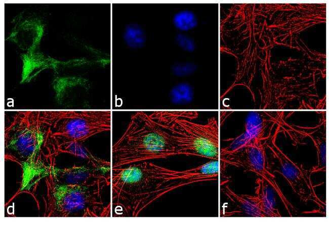
Supportive validation
- Submitted by
- Invitrogen Antibodies (provider)
- Main image

- Experimental details
- For immunofluorescence analysis, retinoic acid (10 uM, 3days) treated SH-SY5Y cells were fixed and permeabilized for detection of Neurofilament-H using Neurofilament-H Recombinant Rabbit Polyclonal Antibody (Product # 711025, 2 µg/mL) and labeled with Goat anti-Rabbit IgG (H+L) Superclonal™ Secondary Antibody, Alexa Fluor® 488 conjugate (Product # A27034, 1:2000). Panel a) shows representative cells that were stained for detection and localization of Neurofilament-H protein (green), Panel b) is stained for nuclei (blue) using SlowFade® Gold Antifade Mountant with DAPI (Product # S36938). Panel c) represents cytoskeletal F-actin staining using Alexa Fluor® 555 Rhodamine Phalloidin (Product # R415, 1:300). Panel d) is a composite image of Panels a, b and c clearly demonstrating cytoplasmic localization of Neurofilament-H. Panel e) shows untreated cells with nuclear localization. Panel f) represents control cells with no primary antibody to assess background.
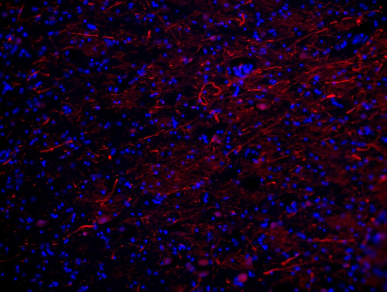
Supportive validation
- Submitted by
- Invitrogen Antibodies (provider)
- Main image

- Experimental details
- Immunofluorescent analysis of the neurofilament heavy chains in frozen sections of human cortex. Tissues were probed with a neurofilament heavy chain monoclonal antibody (Product # 711025, red) at a dilution of 1:250. Tissues were then incubated with Alexa Fluor 594 Goat anti-Mouse IgG (H+L) Secondary Antibody. Nuclei (blue) were stained with DAPI. Data courtesy of Min Sun Kim at Seoul National University, Korea.
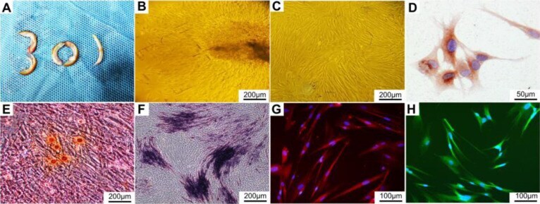
Supportive validation
- Submitted by
- Invitrogen Antibodies (provider)
- Main image

- Experimental details
- Culture and identification of DPSCs. (A) Teeth of New Zealand white rabbits; (B) primary culture; (C) subculture; (D) immunohistochemical staining of vimentin; (E) alizarin red staining; (F) alkaline phosphatase staining; (G) immunofluorescence staining of NF200 (red); (H) immunofluorescence staining of Stro1 (green).