 Explore
 Validate
 Learn
Learn Western blot
 Immunocytochemistry
Antibody data
- Antibody Data
- Antigen structure
- References [0]
- Comments [0]
- Validations
- Western blot [1]
- Other assay [1]
Submit
Validation data
Reference
Comment
Report error
- Product number
- NBP1-05209 - Provider product page

- Provider
- Novus Biologicals
- Proper citation
- Novus Cat#NBP1-05209, RRID:AB_1556328
- Product name
- Mouse Monoclonal NF-H Antibody
- Antibody type
- Monoclonal
- Description
- Affinity purified. Clone AH1 recognizes phosphorylated NF-H KSP sequences. Does not recognize non-phosphorylated KSP sequences.
- Reactivity
- Human, Mouse, Rat, Bovine, Porcine
- Host
- Mouse
- Isotype
- IgG
- Vial size
- 0.1 ml
- Concentration
- 1 mg/ml
- Storage
- Store at 4C short term. Aliquot and store at -20C long term. Avoid freeze-thaw cycles.
No comments: Submit comment
Supportive validation
- Submitted by
- Novus Biologicals (provider)
- Main image

- Experimental details
- Western Blot: NF-H Antibody (AH1) [NBP1-05209] - Analysis of the heavily phosphorylated axonal form of NF-H protein (pNF-H) in neural tissue lysates (20ug/lane) with affinity purified mouse monoclonal anti-pNF-H antibody (NBP1-05209) at dilution of 1:5,000. Lanes on the blot are: [1] Protein size marker, [2] Adult rat whole brain [3] Embryonic (E20) rat whole brain [4] Adult rat spinal cord [5] Adult mouse whole brain [6] Adult mouse spinal cord. Rodent pNF-H protein appears as a single band of about 200kDa in adult rat and mouse lysates, but is not present in early development (Lane 3). Additional bands appearing on the blot (Lane 4) are most likely partially degraded products of pNF-H protein.

Supportive validation
- Submitted by
- Novus Biologicals (provider)
- Main image
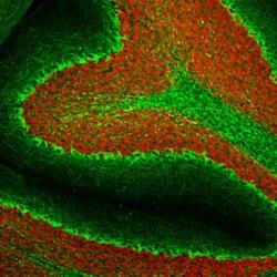
- Experimental details
- Immunohistochemistry Free-Floating: NF-H Antibody (AH1) [NBP1-05209] - Analysis of rat cerebellum section stained with mouse mAb to pNF-H, NBP1-05209, dilution 1:2,000 in green, and costained with rabbit pAb to FOX3/NeuN, dilution 1:5,000 in red. Following transcardial perfusion with 4% paraformaldehyde, brain was post fixed for 24 hours, cut to 45uM, and free-floating sections were stained with above antibodies. The NBP1-05209 antibody stains axons in the granular layer and white matter and prominent basket cell axons surrounding the large Purkinje neurons. The FOX3/NeuN antibody specifically labels nuclei of granular and other neurons, but does not stain Purkinje cells.
