 Explore
 Validate
 Learn
Learn Western blot
Antibody data
- Antibody Data
- Antigen structure
- References [2]
- Comments [0]
- Validations
- Western blot [2]
- Immunohistochemistry [1]
Submit
Validation data
Reference
Comment
Report error
- Product number
- MA3-16722 - Provider product page

- Provider
- Invitrogen Antibodies
- Product name
- NF-H Monoclonal Antibody (NAP4)
- Antibody type
- Monoclonal
- Antigen
- Purifed from natural sources
- Description
- This antibody specifically recognizes the phosphorylated variant of NF-H subunit (~200-220 kDa), showing some weaker reactivity with phosphorylated forms of NF-M.
- Reactivity
- Human, Mouse, Rat, Bovine, Chicken/Avian, Porcine
- Host
- Mouse
- Isotype
- IgG
- Antibody clone number
- NAP4
- Vial size
- 100 µL
- Concentration
- 1 mg/mL
- Storage
- Store at 4°C short term. For long term storage, store at -20°C, avoiding freeze/thaw cycles.
Submitted references Compartmentation of alpha-internexin and neurofilament triplet proteins in cultured hippocampal neurons.
Compartmentation of alpha-internexin and neurofilament triplet proteins in cultured hippocampal neurons.
Benson DL, Mandell JW, Shaw G, Banker G
Journal of neurocytology 1996 Mar;25(3):181-96
Journal of neurocytology 1996 Mar;25(3):181-96
Compartmentation of alpha-internexin and neurofilament triplet proteins in cultured hippocampal neurons.
Benson DL, Mandell JW, Shaw G, Banker G
Journal of neurocytology 1996 Mar;25(3):181-96
Journal of neurocytology 1996 Mar;25(3):181-96
No comments: Submit comment
Supportive validation
- Submitted by
- Invitrogen Antibodies (provider)
- Main image

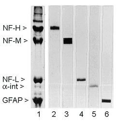
- Experimental details
- Rat spinal cord homogenate showing the major intermediate filament proteins of the nervous system (lane 1). The remaining lanes show blots of this material stainted with various antibodies including 1 (lane 2).
- Submitted by
- Invitrogen Antibodies (provider)
- Main image
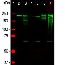
- Experimental details
- Western blot analysis of NF-H in tissue lysates. Samples were incubated in NF-H monoclonal antibody (Product # MA3-16722 using a dilution of 1:10,000. Antibody in green: [1] protein standard (red), [2] rat brain, [3] rat spinal cord, [4] mouse brain, [5] mouse spinal cord, [6] pig spinal cord, [7] cow spinal cord. Strong band at about 200-220 kDa corresponds to the major phosphorylated from of the NF-H subunit. A minor band at about 160 kDa is the non-phosphorylated NF-H. Smaller proteolytic fragments of NF-H are also detected in spinal cord preparations with this antibody.

Supportive validation
- Submitted by
- Invitrogen Antibodies (provider)
- Main image
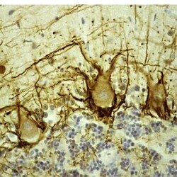
- Experimental details
- Immunohistochemical analysis of NF-H in Paraffin-embedded, formalin-fixed human cerebellar cortex tissue sections. Samples were incubated in NF-H monoclonal antibody (Product # MA3-16722). Staining using the avidin biotin conjugate method. The sections was counterstained with Hematoxylin in blue. This antibody stains prominent basket cell axons surrounding the large Purkinje neurons. Cerebellar granule cell layer is at the bottom of the image, the molecular layer at the top.
