Explore
 Validate
 Learn
Learn Western blot
 ELISA
ELISA Immunocytochemistry
Antibody data
- Antibody Data
- Antigen structure
- References [6]
- Comments [0]
- Validations
- Immunocytochemistry [2]
- Immunohistochemistry [2]
- Other assay [4]
Submit
Validation data
Reference
Comment
Report error
- Product number
- PA1-10002 - Provider product page

- Provider
- Invitrogen Antibodies
- Product name
- NF-H Polyclonal Antibody
- Antibody type
- Polyclonal
- Antigen
- Purifed from natural sources
- Description
- This antibody reacts primarily with the phosphorylated axonal form of NF-H and shows some cross-reactivity with phosphorylated NF-M, which has similar phosphorylation sites to NF-H.
- Reactivity
- Human, Mouse, Rat, Bovine, Canine, Porcine
- Host
- Chicken/Avian
- Isotype
- IgY
- Vial size
- 50 μL
- Concentration
- Conc. Not Determined
- Storage
- 4°C
Submitted references Myelination, axonal loss and Schwann cell characteristics in axonal polyneuropathy compared to controls.
Nano-in-Micro-Particles Consisting of PLGA Nanoparticles Embedded in Chitosan Microparticles via Spray-Drying Enhances Their Uptake in the Olfactory Mucosa.
Epitope-preserving magnified analysis of proteome (eMAP).
Endoplasmic reticulum stress in the dorsal root ganglia regulates large-conductance potassium channels and contributes to pain in a model of multiple sclerosis.
Sensory Neurons of the Dorsal Root Ganglia Become Hyperexcitable in a T-Cell-Mediated MOG-EAE Model of Multiple Sclerosis.
Murine Retinal Citrullination Declines With Age and is Mainly Dependent on Peptidyl Arginine Deiminase 4 (PAD4).
Placheta-Györi E, Brandstetter LM, Zemann-Schälss J, Wolf S, Radtke C
PloS one 2021;16(11):e0259654
PloS one 2021;16(11):e0259654
Nano-in-Micro-Particles Consisting of PLGA Nanoparticles Embedded in Chitosan Microparticles via Spray-Drying Enhances Their Uptake in the Olfactory Mucosa.
Spindler LM, Feuerhake A, Ladel S, Günday C, Flamm J, Günday-Türeli N, Türeli E, Tovar GEM, Schindowski K, Gruber-Traub C
Frontiers in pharmacology 2021;12:732954
Frontiers in pharmacology 2021;12:732954
Epitope-preserving magnified analysis of proteome (eMAP).
Park J, Khan S, Yun DH, Ku T, Villa KL, Lee JE, Zhang Q, Park J, Feng G, Nedivi E, Chung K
Science advances 2021 Nov 12;7(46):eabf6589
Science advances 2021 Nov 12;7(46):eabf6589
Endoplasmic reticulum stress in the dorsal root ganglia regulates large-conductance potassium channels and contributes to pain in a model of multiple sclerosis.
Yousuf MS, Samtleben S, Lamothe SM, Friedman TN, Catuneanu A, Thorburn K, Desai M, Tenorio G, Schenk GJ, Ballanyi K, Kurata HT, Simmen T, Kerr BJ
FASEB journal : official publication of the Federation of American Societies for Experimental Biology 2020 Sep;34(9):12577-12598
FASEB journal : official publication of the Federation of American Societies for Experimental Biology 2020 Sep;34(9):12577-12598
Sensory Neurons of the Dorsal Root Ganglia Become Hyperexcitable in a T-Cell-Mediated MOG-EAE Model of Multiple Sclerosis.
Yousuf MS, Noh MC, Friedman TN, Zubkow K, Johnson JC, Tenorio G, Kurata HT, Smith PA, Kerr BJ
eNeuro 2019 Mar-Apr;6(2)
eNeuro 2019 Mar-Apr;6(2)
Murine Retinal Citrullination Declines With Age and is Mainly Dependent on Peptidyl Arginine Deiminase 4 (PAD4).
Hollingsworth TJ, Radic MZ, Beranova-Giorgianni S, Giorgianni F, Wang Y, Iannaccone A
Investigative ophthalmology & visual science 2018 Aug 1;59(10):3808-3815
Investigative ophthalmology & visual science 2018 Aug 1;59(10):3808-3815
No comments: Submit comment
Supportive validation
- Submitted by
- Invitrogen Antibodies (provider)
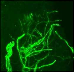
- Main image
- Experimental details
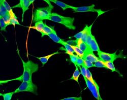
- Immunofluorescent analysis of NEFH using a polyclonal antibody (Product # PA1-10002).
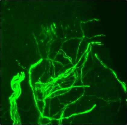
- Submitted by
- Invitrogen Antibodies (provider)
- Main image
- Experimental details
- Immunofluorescent analysis of NEFH using a polyclonal antibody (Product # PA1-10002).

Supportive validation
- Submitted by
- Invitrogen Antibodies (provider)
- Main image
- Experimental details
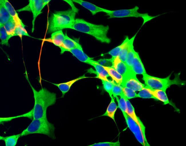
- Immunohistochemical analysis of human skin stained with Neurofilament, Heavy chain Polyclonal Antibody (Product # PA1-10002). Free-floating, formalin-fixed frozen human skin tissue sections (60 µm) were stained with PA1-10002 (1:2000) followed by a fluorescently-conjugated donkey anti-chicken IgY secondary antibody. Tissues were visualized by confocal microscopy. Data courtesy of the Innovators Program.
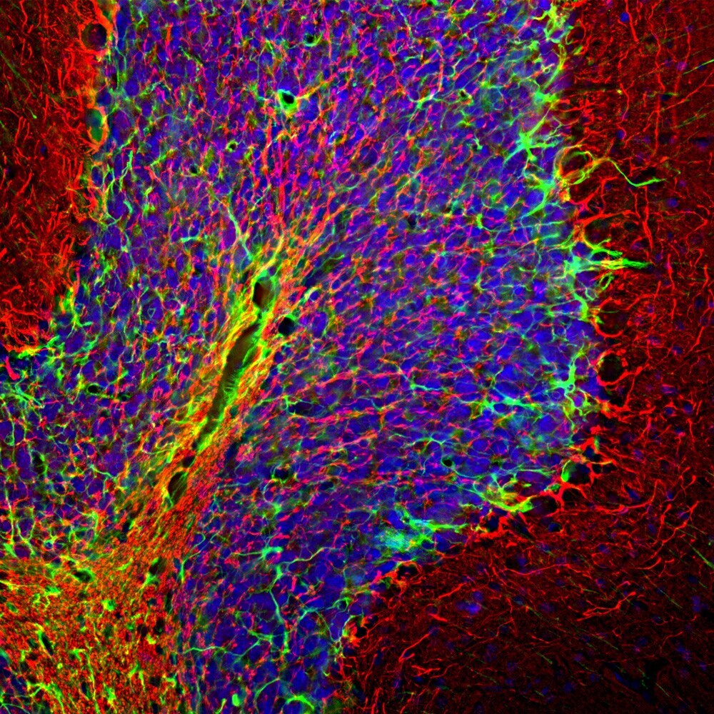
- Submitted by
- Invitrogen Antibodies (provider)
- Main image
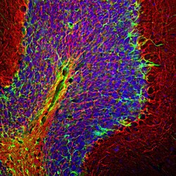
- Experimental details
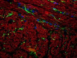
- Immunohistological analysis of NR-H in rat cerebellum. The rat cerebellum section was obtained following transcardial perfusion of the rat with 4% paraformaldehyde, brain was post fixed for 24 hours, and cut to 45µM. Free-floating sections were stained with an NF-H polyclonal antibody (Product # PA1-10002) at a dilution of 1:5,000 as seen in red, and costained with a GFAP polyclonal antibody (Product # PA1-10019) at a dilution of 1:5,000 as seen in green, and with DAPI staining the nuclear DNA in blue. The NF-H antibody labels network of axons of different neurons, while the GFAP antibody stains astrocytes and other glial cells.
Supportive validation
- Submitted by
- Invitrogen Antibodies (provider)
- Main image
- Experimental details
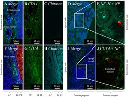
- FIGURE 5 Permeation of PLGA nanoparticles (520 nm; red) (A-E) and PLGA-chitosan nano-in-micro particles (NiMPs) (F-J) . Maximum intensity projection (MIP) of Confocal Laser Scanning Microscopy (CLSM) images stitched together from a z-stack, taken 15 min after application to the apical side of porcine olfactory mucosa. Cell nuclei stained with DAPI (blue). Red arrows mark nanoparticles (red) within the tissue. CD14 (green) labelled with anti-mouse Alexa Fluor (r) 647 secondary antibody (A, B, F, G) . NF-H (cyan) labelled with anti-chicken FITC secondary antibody (D, E) . Chitosan labeled with fluorescein (cyan) (H) . Overlapping signals of red and green appear yellow (F) due to co-localization of PLGA nanoparticles (red) and chitosan (green). PLGA nanoparticles (single red channel) displayed in Supplementary Figure S6B, C . LP, Lamina propria; BL, Basal cell layer; PL, Particle layer; EL, Epithelial cell layer.
- Submitted by
- Invitrogen Antibodies (provider)
- Main image
- Experimental details
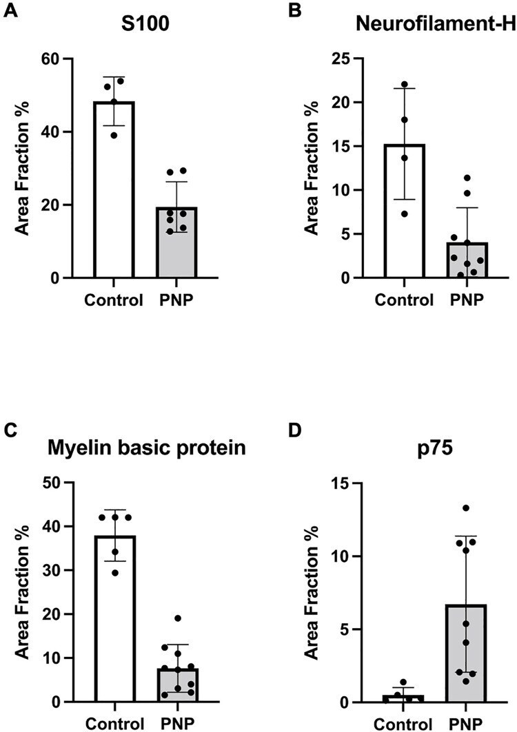
- 10.1371/journal.pone.0259654.g002 Fig 2 Immunohistochemistry of nerve sections. Area fraction of immunosignals in nerve biopsies obtained from PNP patients compared to controls. S100 positive Schwann cells (A) were significantly reduced in PNP. Neurofilament-H (B) was highly significantly lower in the PNP group. There was reduced immunoreactivity to the myelination parameter myelin basic protein in PNP patients (C), while immature p75-positive Schwann cells were increased (D). PNP : Polyneuropathy .
- Submitted by
- Invitrogen Antibodies (provider)
- Main image
- Experimental details
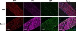
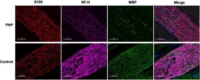
- Fig 3 Immunofluorescent staining. S100 positive Schwann cells (red), neurofilament-H (NF-H; purple) and myelin basic protein (MBP; green) are reduced in polyneuropathy (PNP). In the merged panel, cell bodies are counter-stained with DAPI (blue). Imaged at 20x magnification under equal exposure times; scale bar: 100 mum.
- Submitted by
- Invitrogen Antibodies (provider)
- Main image
- Experimental details
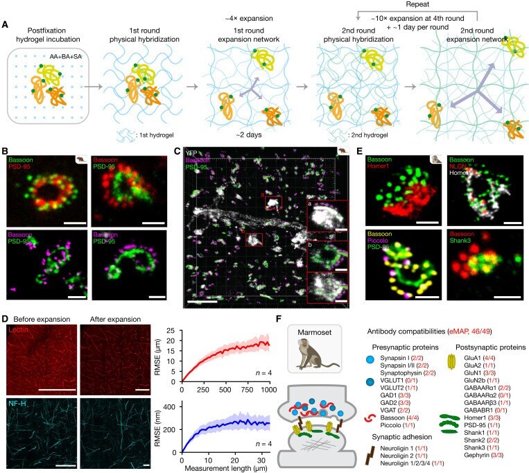
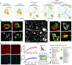
- Fig. 2. eMAP offers high expansion factors by recursive embedding while preserving the spatial organization of molecules. ( A ) Schematic of the recursive embedding of an eMAP-processed tissue. ( B ) Individual synapses in eMAP-processed, 10x linearly expanded mouse brain tissues visualized by different sets of Bassoon and PSD-95 antibodies: Bassoon SYSY 141004 and PSD-95 NeuroMab 75-028 (top), and Bassoon SYSY 141003 and PSD-95 NeuroMab 75-028 (bottom). Scale bars, 200 nm (2 mum in expanded). ( C ) 3D-rendered image of a 50-mum-thick (5 mum before expansion) mouse brain slice stained with anti-YFP (white), Bassoon (magenta), and PSD-95 (green) antibodies. Scale bars, 500 nm (5 mum in expanded). (A) and (B) are inset images of respective dendritic spines highlighted in the 3D-rendered image. Scale bars, 200 nm (2 mum in expanded). ( D ) Images showing Lectin-stained (top) and NF-H-stained (bottom) tissues before and after expansion. Scale bars, 500 mum (Lectin) and 50 mum (NF-H). The plots on the right show the root mean square error (RMSE) at different measurement lengths mesoscale (top) and microscale (bottom). ( E ) Individual synapses in eMAP-processed, 10x linearly expanded marmoset brain tissues visualized by different sets of antibodies. Scale bars, 200 nm (2 mum in expanded). ( F ) Antibody compatibility in eMAP-processed marmoset brain tissue for the 27 SyPs in Fig. 1D .