Explore
 Validate
 Learn
Learn Western blot
 ELISA
ELISAAntibody data
- Antibody Data
- Antigen structure
- References [0]
- Comments [0]
- Validations
- Western blot [4]
- Immunohistochemistry [1]
Submit
Validation data
Reference
Comment
Report error
- Product number
- PA3-16721 - Provider product page

- Provider
- Invitrogen Antibodies
- Product name
- NF-H Polyclonal Antibody
- Antibody type
- Polyclonal
- Antigen
- Purifed from natural sources
- Reactivity
- Human, Mouse, Rat, Bovine, Porcine
- Host
- Rabbit
- Isotype
- IgG
- Vial size
- 50 µL
- Concentration
- Conc. Not Determined
- Storage
- Store at 4°C short term. For long term storage, store at -20°C, avoiding freeze/thaw cycles.
No comments: Submit comment
Supportive validation
- Submitted by
- Invitrogen Antibodies (provider)
- Main image

- Experimental details
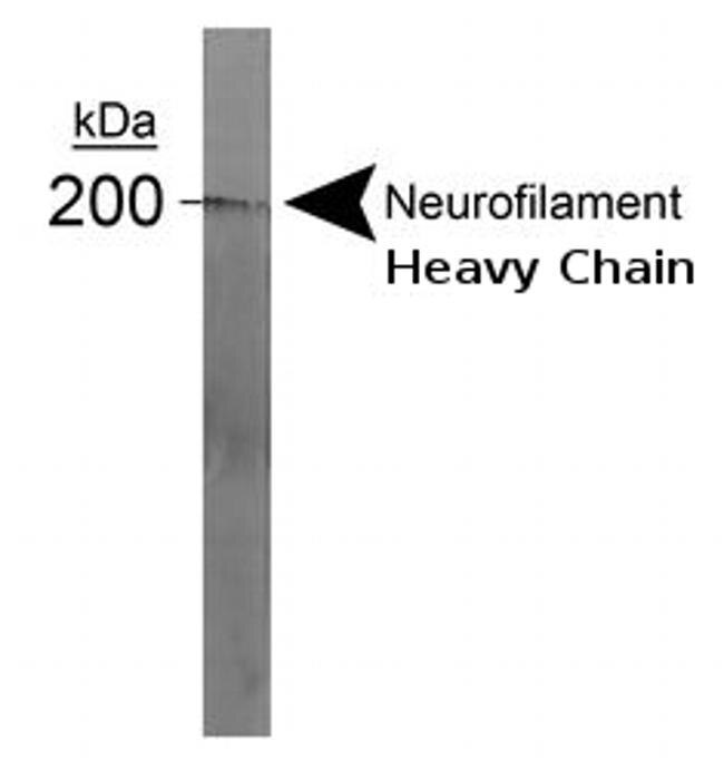
- Strip blot of crude rat spinal extract stained with Neurofilament Heavy Chain antibody (clone DA2) (Product # PA3-16721). A prominent band appears at 200kDa which corresponds to phosphorylated NF-H.
- Submitted by
- Invitrogen Antibodies (provider)
- Main image

- Experimental details
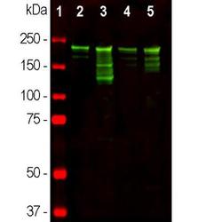
- Western blot analysis of NF-H in Different tissue lysates. Samples were incubated in NF-H polyclonal antibody (Product # PA3-16721 using a dilution of 1:10,000. Antibody in green: [1] protein standard (red), [2] rat brain, [3] rat spinal cord [4] mouse brain, and [5] mouse spinal cord lysate. Strong band at about 220 kDa corresponds to the phosphorylated axonal form of the NF-H subunit. Smaller proteolytic fragments of NF-H are also detected with RPCA-NF-H antibody.

- Submitted by
- Invitrogen Antibodies (provider)
- Main image
- Experimental details
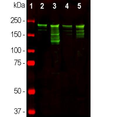
- Western blot analysis of NF-H in Different tissue lysates. Samples were incubated in NF-H polyclonal antibody (Product # PA3-16721 using a dilution of 1:10,000. Antibody in green: [1] protein standard (red), [2] rat brain, [3] rat spinal cord [4] mouse brain, and [5] mouse spinal cord lysate. Strong band at about 220 kDa corresponds to the phosphorylated axonal form of the NF-H subunit. Smaller proteolytic fragments of NF-H are also detected with RPCA-NF-H antibody.
- Submitted by
- Invitrogen Antibodies (provider)
- Main image
- Experimental details
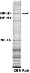
- Western blot analysis of NF-H in 200 kDa Neurofilament Heavy expression in rat spinal cord extract. Samples were incubated in NF-H polyclonal antibody (Product # PA3-16721). Lane 1: Coomassie Brilliant Blue stained; Lane 2: probed with rabbit anti-Neurofilament Heavy antibody. The NF-H corresponds to a weight of 200 kDa.

Supportive validation
- Submitted by
- Invitrogen Antibodies (provider)
- Main image

- Experimental details
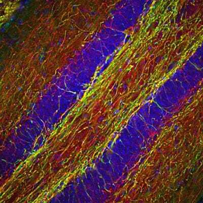
- Immunohistochemical analysis of NF-H in Mouse hippocampus section. Samples were incubated in NF-H polyclonal antibody (Product # PA3-16721) using a dilution of 1:2000. Antibody in red, and costained with mouse mAb to myelin basic protein (MBP), dilution 1:5,000 in green. The blue is DAPI staining of nuclear DNA. Following transcardial perfusion with 4% paraformaldehyde, brain was post fixed for 24 hours, cut to 45 µM, and free-floating sections were stained with above antibodies. The NF-H antibody labels a network of axons of different neurons, while the MBP antibody stains myelin sheath around these axons.