Explore
 Validate
 Learn
Learn Western blot
 ELISA
ELISA Immunocytochemistry
Antibody data
- Antibody Data
- Antigen structure
- References [2]
- Comments [0]
- Validations
- Immunocytochemistry [6]
- Immunohistochemistry [1]
- Other assay [1]
Submit
Validation data
Reference
Comment
Report error
- Product number
- PA3-16753 - Provider product page

- Provider
- Invitrogen Antibodies
- Product name
- NF-H Polyclonal Antibody
- Antibody type
- Polyclonal
- Antigen
- Purifed from natural sources
- Reactivity
- Human, Mouse, Rat, Bovine, Canine, Feline, Porcine
- Host
- Chicken/Avian
- Isotype
- IgY
- Vial size
- 50 μL
- Concentration
- Conc. Not Determined
- Storage
- Store at 4°C short term. For long term storage, store at -20°C, avoiding freeze/thaw cycles.
Submitted references SLC13A5/sodium-citrate co-transporter overexpression causes disrupted white matter integrity and an autistic-like phenotype.
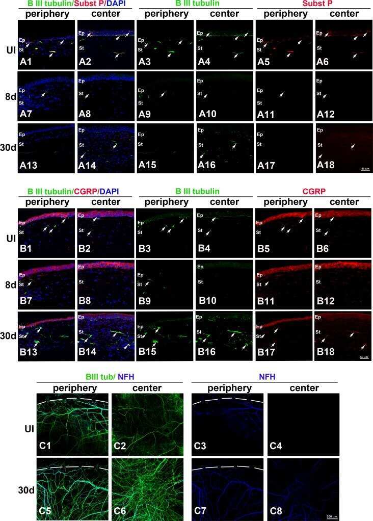
Degeneration and regeneration of corneal nerves in response to HSV-1 infection.
Rigby MJ, Orefice NS, Lawton AJ, Ma M, Shapiro SL, Yi SY, Dieterich IA, Frelka A, Miles HN, Pearce RA, Yu JPJ, Li L, Denu JM, Puglielli L
Brain communications 2022 Feb;4(1):fcac002
Brain communications 2022 Feb;4(1):fcac002
Degeneration and regeneration of corneal nerves in response to HSV-1 infection.
Chucair-Elliott AJ, Zheng M, Carr DJ
Investigative ophthalmology & visual science 2015 Jan 13;56(2):1097-107
Investigative ophthalmology & visual science 2015 Jan 13;56(2):1097-107
No comments: Submit comment
Supportive validation
- Submitted by
- Invitrogen Antibodies (provider)
- Main image
- Experimental details
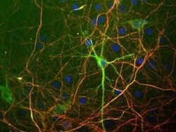
- Immunocytochemistry analysis of NF-H in mixed rat neuron/glial cultures. Samples were incubated in NF-H polyclonal antibody (Product # PA3-16753). Neurofilament Light (NF-L) antibody [green] and Neurofilament Heavy (NF-H) antibody. Blue is a DNA stain. NF-H antibody binds primarily to the phosphorylated axonal forms of NF-H, in contrast to NF-L antibody which stains both axonal and dendritic/perikaryal neurofilaments. The surrounding axonal profiles are orange due to staining of both the NF-H and NF-L antibodies.

- Submitted by
- Invitrogen Antibodies (provider)
- Main image

- Experimental details
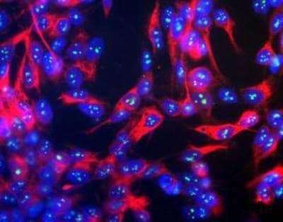
- Immunocytochemistry analysis of NF-H in SH-SY5Y cells. Samples were incubated in NF-H polyclonal antibody (Product # PA3-16753). Neurofilament Heavy Antibody (red) and Fibrillarin Antibody (green). Nuclear DNA is stained with Hoechst dye (blue).

- Submitted by
- Invitrogen Antibodies (provider)
- Main image

- Experimental details
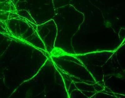
- Immunocytochemistry analysis of NF-H in Rat neurons. Samples were incubated in NF-H polyclonal antibody (Product # PA3-16753).
- Submitted by
- Invitrogen Antibodies (provider)
- Main image

- Experimental details
- Immunocytochemistry analysis of NF-H in mixed rat neuron/glial cultures. Samples were incubated in NF-H polyclonal antibody (Product # PA3-16753). Neurofilament Light (NF-L) antibody [green] and Neurofilament Heavy (NF-H) antibody. Blue is a DNA stain. NF-H antibody binds primarily to the phosphorylated axonal forms of NF-H, in contrast to NF-L antibody which stains both axonal and dendritic/perikaryal neurofilaments. The surrounding axonal profiles are orange due to staining of both the NF-H and NF-L antibodies.

- Submitted by
- Invitrogen Antibodies (provider)
- Main image

- Experimental details
- Immunocytochemistry analysis of NF-H in SH-SY5Y cells. Samples were incubated in NF-H polyclonal antibody (Product # PA3-16753). Neurofilament Heavy Antibody (red) and Fibrillarin Antibody (green). Nuclear DNA is stained with Hoechst dye (blue).
- Submitted by
- Invitrogen Antibodies (provider)
- Main image

- Experimental details
- Immunocytochemistry analysis of NF-H in Rat neurons. Samples were incubated in NF-H polyclonal antibody (Product # PA3-16753).

Supportive validation
- Submitted by
- Invitrogen Antibodies (provider)
- Main image
- Experimental details
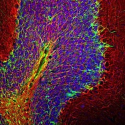
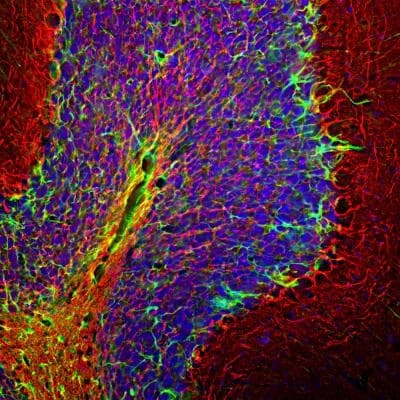
- Immunohistochemical analysis of NF-H in rat cerebellum sections. Samples were incubated in NF-H polyclonal antibody (Product # PA3-16753) using a dilution of 1:5,000 (Red). Costained with rabbit GFAP pAb, dilution 1:5,000 (Green). DAPI staining of nuclear DNA (Blue). Following transcardial perfusion with 4% paraformaldehyde, brain was post fixed for 24 hrs, cut to 45 µM, and free floating sections were stained with above antibodies. The NF-H antibody labels network of axons of different neurons, while the GFAP antibody stains astrocytes and other glial cells.
Supportive validation
- Submitted by
- Invitrogen Antibodies (provider)
- Main image

- Experimental details
- NULL