Explore
 Validate
 Learn
Learn Western blot
 Immunocytochemistry
Antibody data
- Antibody Data
- Antigen structure
- References [6]
- Comments [0]
- Validations
- Immunocytochemistry [6]
- Immunohistochemistry [1]
- Flow cytometry [2]
- Other assay [4]
Submit
Validation data
Reference
Comment
Report error
- Product number
- MA3-16771 - Provider product page

- Provider
- Invitrogen Antibodies
- Product name
- Fibrillarin Monoclonal Antibody (38F3)
- Antibody type
- Monoclonal
- Antigen
- Other
- Description
- Suggested positive control: antigen standard for FBL (transient overexpression lysate).
- Reactivity
- Human, Mouse, Rat, Bovine, Chicken/Avian, Drosophila, Porcine, Yeast
- Host
- Mouse
- Isotype
- IgG
- Antibody clone number
- 38F3
- Vial size
- 250 μL
- Concentration
- Conc. not determined
- Storage
- Store at 4°C short term. For long term storage, store at -20°C, avoiding freeze/thaw cycles.
Submitted references Complete loss of H3K9 methylation dissolves mouse heterochromatin organization.
H19-Dependent Transcriptional Regulation of β3 and β4 Integrins Upon Estrogen and Hypoxia Favors Metastatic Potential in Prostate Cancer.
Optimized protocol for combined PALM-dSTORM imaging.
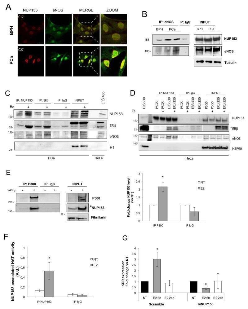
Nucleoporin 153 regulates estrogen-dependent nuclear translocation of endothelial nitric oxide synthase and estrogen receptor beta in prostate cancer.
Olfactory memory is enhanced in mice exposed to extremely low-frequency electromagnetic fields via Wnt/β-catenin dependent modulation of subventricular zone neurogenesis.
40S ribosome biogenesis co-factors are essential for gametophyte and embryo development.
Montavon T, Shukeir N, Erikson G, Engist B, Onishi-Seebacher M, Ryan D, Musa Y, Mittler G, Meyer AG, Genoud C, Jenuwein T
Nature communications 2021 Jul 16;12(1):4359
Nature communications 2021 Jul 16;12(1):4359
H19-Dependent Transcriptional Regulation of β3 and β4 Integrins Upon Estrogen and Hypoxia Favors Metastatic Potential in Prostate Cancer.
Bacci L, Aiello A, Ripoli C, Loria R, Pugliese D, Pierconti F, Rotili D, Strigari L, Pinto F, Bassi PF, Mai A, Grassi C, Pontecorvi A, Falcioni R, Farsetti A, Nanni S
International journal of molecular sciences 2019 Aug 17;20(16)
International journal of molecular sciences 2019 Aug 17;20(16)
Optimized protocol for combined PALM-dSTORM imaging.
Glushonkov O, Réal E, Boutant E, Mély Y, Didier P
Scientific reports 2018 Jun 8;8(1):8749
Scientific reports 2018 Jun 8;8(1):8749
Nucleoporin 153 regulates estrogen-dependent nuclear translocation of endothelial nitric oxide synthase and estrogen receptor beta in prostate cancer.
Re A, Colussi C, Nanni S, Aiello A, Bacci L, Grassi C, Pontecorvi A, Farsetti A
Oncotarget 2018 Jun 15;9(46):27985-27997
Oncotarget 2018 Jun 15;9(46):27985-27997
Olfactory memory is enhanced in mice exposed to extremely low-frequency electromagnetic fields via Wnt/β-catenin dependent modulation of subventricular zone neurogenesis.
Mastrodonato A, Barbati SA, Leone L, Colussi C, Gironi K, Rinaudo M, Piacentini R, Denny CA, Grassi C
Scientific reports 2018 Jan 10;8(1):262
Scientific reports 2018 Jan 10;8(1):262
40S ribosome biogenesis co-factors are essential for gametophyte and embryo development.
Missbach S, Weis BL, Martin R, Simm S, Bohnsack MT, Schleiff E
PloS one 2013;8(1):e54084
PloS one 2013;8(1):e54084
No comments: Submit comment
Supportive validation
- Submitted by
- Invitrogen Antibodies (provider)
- Main image

- Experimental details
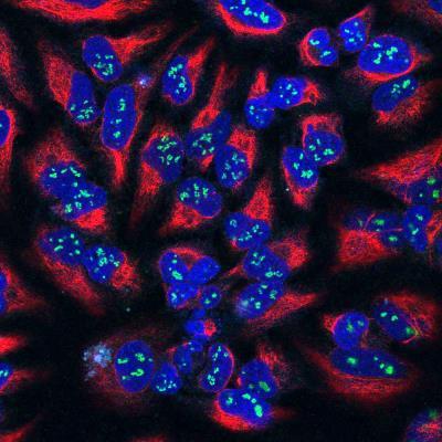
- Immunocytochemistry analysis of Fibrillarin in HeLa cells. Samples were incubated in Fibrillarin monoclonal antibody (Product # MA3-16771) using a dilution of 1:100. Antibody from concentrated tissue culture media (Green), and costained with chicken vimentin pAb (Red), dilution 1:1,000. Nuclear DNA is revealed with the DAPI stain (Blue). The fibrillarin antibody shows strong staining of nucleoli in the nucleus, while the vimentin antibody reveals cytoplasmic intermediate filaments.
- Submitted by
- Invitrogen Antibodies (provider)
- Main image

- Experimental details
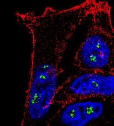
- Immunocytochemistry analysis of Fibrillarin in MCF7 cells. Samples were incubated in Fibrillarin monoclonal antibody (Product # MA3-16771) followed by Alexa Fluor 488-conjugated Goat to mouse IgG secondary antibody (green). Actin filaments were labeled with Alexa Fluor 568 phalloidin (red). DAPI was used to stain the cell nuclei (blue).

- Submitted by
- Invitrogen Antibodies (provider)
- Main image

- Experimental details
- Immunofluorescence analysis of Fibrillarin was performed using 70% confluent log phase HeLa cells. The cells were fixed with 4% paraformaldehyde for 10 minutes, permeabilized with 0.1% Triton™ X-100 for 15 minutes, and blocked with 1% BSA for 1 hour at room temperature. The cells were labeled with Fibrillarin Monoclonal Antibody (38F3) (Product # MA3-16771) at 1:250 dilution in 0.1% BSA, incubated at 4 degree Celsius overnight and then labeled with Goat anti-Mouse IgG (H+L) Superclonal™ Secondary Antibody, Alexa Fluor® 488 conjugate (Product # A28175) for 45 minutes at room temperature (Panel a: green). Nuclei (Panel b: blue) were stained with ProLong™ Diamond Antifade Mountant with DAPI (Product # P36962). F-actin (Panel c: red) was stained with Rhodamine Phalloidin (Product # R415, 1:300). Panel d represents the merged image showing nucleolar localization. Panel e represents control cells with no primary antibody to assess background. The images were captured at 60X magnification.

- Submitted by
- Invitrogen Antibodies (provider)
- Main image

- Experimental details
- Immunocytochemistry analysis of Fibrillarin in HeLa cells. Samples were incubated in Fibrillarin monoclonal antibody (Product # MA3-16771) using a dilution of 1:100. Antibody from concentrated tissue culture media (Green), and costained with chicken vimentin pAb (Red), dilution 1:1,000. Nuclear DNA is revealed with the DAPI stain (Blue). The fibrillarin antibody shows strong staining of nucleoli in the nucleus, while the vimentin antibody reveals cytoplasmic intermediate filaments.

- Submitted by
- Invitrogen Antibodies (provider)
- Main image
- Experimental details
- Immunocytochemistry analysis of Fibrillarin in MCF7 cells. Samples were incubated in Fibrillarin monoclonal antibody (Product # MA3-16771) followed by Alexa Fluor 488-conjugated Goat to mouse IgG secondary antibody (green). Actin filaments were labeled with Alexa Fluor 568 phalloidin (red). DAPI was used to stain the cell nuclei (blue).

- Submitted by
- Invitrogen Antibodies (provider)
- Main image

- Experimental details
- Immunofluorescence analysis of Fibrillarin was performed using 70% confluent log phase HeLa cells. The cells were fixed with 4% paraformaldehyde for 10 minutes, permeabilized with 0.1% Triton™ X-100 for 15 minutes, and blocked with 1% BSA for 1 hour at room temperature. The cells were labeled with Fibrillarin Monoclonal Antibody (38F3) (Product # MA3-16771) at 1:250 dilution in 0.1% BSA, incubated at 4 degree Celsius overnight and then labeled with Goat anti-Mouse IgG (H+L) Superclonal™ Secondary Antibody, Alexa Fluor® 488 conjugate (Product # A28175) for 45 minutes at room temperature (Panel a: green). Nuclei (Panel b: blue) were stained with ProLong™ Diamond Antifade Mountant with DAPI (Product # P36962). F-actin (Panel c: red) was stained with Rhodamine Phalloidin (Product # R415, 1:300). Panel d represents the merged image showing nucleolar localization. Panel e represents control cells with no primary antibody to assess background. The images were captured at 60X magnification.

Supportive validation
- Submitted by
- Invitrogen Antibodies (provider)
- Main image

- Experimental details
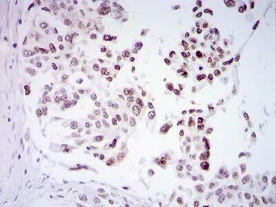
- Immunohistochemical analysis of Fibrillarin in human ovarian cancer. Samples were incubated in Fibrillarin monoclonal antibody (Product # MA3-16771) followed by DAB with hematoxylin counterstain.
Supportive validation
- Submitted by
- Invitrogen Antibodies (provider)
- Main image

- Experimental details
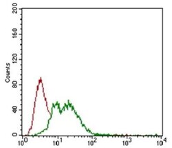
- Flow cytometry of Fibrillarin in HEK293 cells. Samples were incubated in Fibrillarin monoclonal antibody (Product # MA3-16771) using a dilution of 1:400 followed by Alexa Fluor 488 secondary. Secondary (shown in green) alongside unstained cells (shown in red).

- Submitted by
- Invitrogen Antibodies (provider)
- Main image
- Experimental details
- Flow cytometry of Fibrillarin in HEK293 cells. Samples were incubated in Fibrillarin monoclonal antibody (Product # MA3-16771) using a dilution of 1:400 followed by Alexa Fluor 488 secondary. Secondary (shown in green) alongside unstained cells (shown in red).

Supportive validation
- Submitted by
- Invitrogen Antibodies (provider)
- Main image
- Experimental details
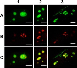
- Figure 1 Confocal microscopy imaging of nucleolar sub-domains in HeLa cells. Column 1: NPM protein fused to the fluorescent protein eGFP was overexpressed after cell transient transfection in order to image the nucleoli granular component ( A1 ). Endogenous RPA was immunolabelled with A647-Ab ( B1 ). The merged images evidenced the specificity of the labelling strategy ( C1 ). Column 2: eGFP-NPM localization is displayed in panel A2. Endogenous Fib was immunolabelled with A647-Ab ( B2 ). The merged images evidenced the spatial overlap between the two domains ( C2 ). Column 3: Endogenous Fib was immunolabelled with A555-Ab ( A3 ). Endogenous RPA was immunolabelled with A647-Ab ( B3 ). The merged images evidenced the spatial overlap between the two domains ( C3 ). Scale bar: 5 um.

- Submitted by
- Invitrogen Antibodies (provider)
- Main image
- Experimental details
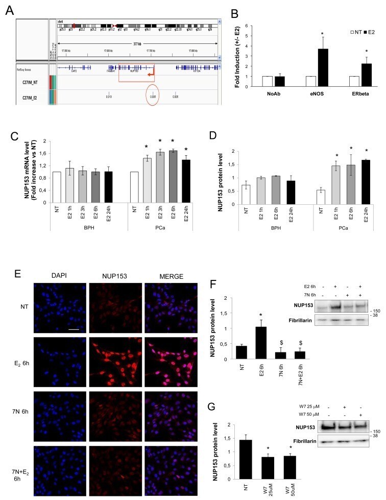
- Figure 2 eNOS binds Nup153 promoter and regulates its expression in an estrogen-dependent manner (A) Integrated Genome Viewer (IGV 2.3) screenshot showing peaks of eNOS identified by ChIP-Seq at the genomic regions encoding Nup153 in PCa cells (C27IM) in the absence (NT) or presence of 17beta-estradiol (E 2 , 10 -7 M, 45min). Region amplified in panel B is indicated with a red circle. (B) Recruitment of eNOS and ERbeta on the promoter region of Nup153 by ChIPs in the presence or absence of E 2 (45 min) in C27IM cells. The immunoprecipitations were performed using anti-eNOS (Type III, BD), anti-ERbeta (L-20) or no antibody (NoAb) as a negative control. Values are represented as fold of induction (+/-E 2 ) and as mean +/-SEM of 3 independent experiments. * p
- Submitted by
- Invitrogen Antibodies (provider)
- Main image
- Experimental details
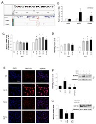
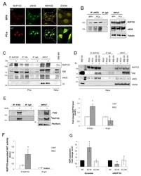
- Figure 3 Nup153 forms complexes with eNOS, ERbeta and p300 (A) Confocal analysis of Prostate Cancer (PCa, C27IM) and Benign Prostatic Hyperplasia (BPH, C17IM) cells stained with antibodies to Nup153 ([QE5]; red) or eNOS (Type III; green). Scale bar: 20mum (Nup153, eNOS and MERGE; zoomed area is showed). (B) Analysis of Nup153 interaction with eNOS by Co-Immunoprecipitation in PCa and BPH cells in basal condition. Immunoprecipitation with IgG served as negative control. (C) Analysis of Nup153 interaction with eNOS and ERbeta by Co-Immunoprecipitation in PCa cells in basal condition or upon E 2 treatment (10 -7 M, 24h). PCa cells were immunoprecipitated with Nup153 Antibody ([7AB], Abcam) or with ERbeta (GeneTex, #110607). Immunoprecipitation with IgG served as negative control. Membranes were blotted with specific antibodies to Nup153 ([Q5], Abcam), ERbeta (GeneTex, #110607) and eNOS (Type III, BD). H1 served as control. 4mug of transfected HeLa with human ERbeta short isoform (485aa) were used as positive control for Estrogen Receptor. Total proteins were resolved by SDS-PAGE using a 10% Invitrogen precast gel (NuPage and MES buffer). (D) Analysis of Nup153 interaction with eNOS and ERbeta by Co-Immunoprecipitation in HeLa cells transfected with empty vector (PSG5) or with human ERbeta full-length (ERbeta 530aa), before and after E 2 treatment (10 -7 M, 24h). HeLa cells were immunoprecipitated with Nup153 Antibody ([7AB], Abcam). Immunoprecipitation with IgG served as negativ
- Submitted by
- Invitrogen Antibodies (provider)
- Main image
- Experimental details
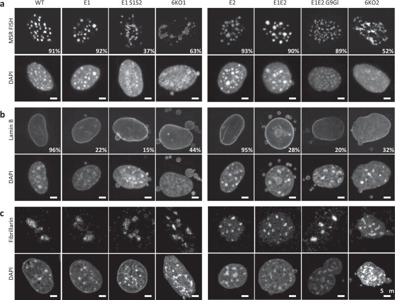
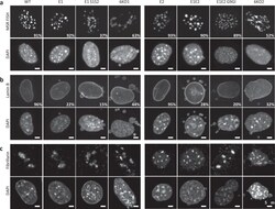
- Fig. 6 Collapse of heterochromatin in compound H3K9 KMT-mutant MEF cells. a DNA FISH for major satellite repeats (MSR) in control and compound H3K9 KMT mutant. Percentages indicate cells that display the shown pattern. N = 501 cells examined for WT, 303 for E1 , 352 for E1 S1S2 , 304 for 6KO1 , 308 for E2 , 311 for E1E2 , 301 for E1E2 G9Gl , and 309 for 6KO2 over three independent experiments. For each image, lower panels show DAPI counterstaining of the same nuclei. b Immunofluorescence for Lamin B, marking the nuclear lamina. Percentages indicate cells that display the shown pattern. N = 680 cells examined for WT, 785 for E1 , 302 for E1 S1S2 , 305 for 6KO1 , 573 for E2 , 499 for E1E2 , 558 for E1E2 G9Gl, and 427 for 6KO2 over three independent experiments. For each image, lower panels show DAPI counterstaining of the same nuclei. c Immunofluorescence for Fibrillarin, marking nucleoli. Representative images ( N = 100 cells) are shown. For each image, lower panels show DAPI counterstaining of the same nuclei. Scale bars represent 5 mum.