Antibody data
- Antibody Data
- Antigen structure
- References [3]
- Comments [0]
- Validations
- Western blot [13]
- Immunocytochemistry [1]
- Immunohistochemistry [2]
- Other assay [1]
Submit
Validation data
Reference
Comment
Report error
- Product number
- PA5-29801 - Provider product page
- Provider
- Invitrogen Antibodies
- Product name
- Fibrillarin Polyclonal Antibody
- Antibody type
- Polyclonal
- Antigen
- Recombinant protein fragment
- Description
- Recommended positive controls: 293T, H1299, Molt-4, Neuro2A, PC-12, A549, A549 nuclear extract, Jurkat, Jurkat nuclear extract, SK-N-SH, SK-N-SH nuclear extract, IMR32, IMR32 nuclear extract.
- Concentration
- 0.36 mg/mL
Submitted references Nucleolar localization of the ErbB3 receptor as a new target in glioblastoma.
9-Cyanopyronin probe palette for super-multiplexed vibrational imaging.
Auxiliary genetic analysis in a Chinese adolescent NPH family by single nucleotide polymorphism screening.
Tagliaferro M, Rosa P, Bellenchi GC, Bastianelli D, Trotta R, Tito C, Fazi F, Calogero A, Ponti D
BMC molecular and cell biology 2022 Mar 7;23(1):13
9-Cyanopyronin probe palette for super-multiplexed vibrational imaging.
Miao Y, Qian N, Shi L, Hu F, Min W
Nature communications 2021 Jul 26;12(1):4518
Auxiliary genetic analysis in a Chinese adolescent NPH family by single nucleotide polymorphism screening.
Tang C, Zhou D, Tan R, Zhong X, Xiao X, Qin D, Liu Y, Hu J, Liu Y
Molecular medicine reports 2020 Mar;21(3):1115-1124
No comments: Submit comment
Supportive validation
- Submitted by
- Invitrogen Antibodies (provider)
- Main image
- Experimental details
- Western blot analysis of Fibrillarin using whole cell lysate (30 µg) and nuclear extract (30 µg) from A549 cells. Samples were loaded onto a 10% SDS-PAGE gel and probed with a Fibrillarin polyclonal antibody (Product # PA5-29801) at a dilution of 1:500.
- Submitted by
- Invitrogen Antibodies (provider)
- Main image
- Experimental details
- Western Blot using Fibrillarin Polyclonal Antibody (Product # PA5-29801). Sample (30 µg of whole cell lysate). Lane A: 293T. Lane B: H1299. Lane C: Molt-4. 12% SDS PAGE. Fibrillarin Polyclonal Antibody (Product # PA5-29801) diluted at 1:1,000.
- Submitted by
- Invitrogen Antibodies (provider)
- Main image
- Experimental details
- Fibrillarin Polyclonal Antibody detects Fibrillarin protein by western blot analysis. Whole cell lysate (30 µg) and nuclear extract (30 µg) from SK-N-SH cells.10 % SDS-PAGE. Fibrillarin Polyclonal Antibody (Product # PA5-29801) dilution: 1:500.
- Submitted by
- Invitrogen Antibodies (provider)
- Main image
- Experimental details
- Fibrillarin Polyclonal Antibody detects Fibrillarin protein by western blot analysis. Whole cell lysate (30 µg) and nuclear extract (30 µg) from A549 cells.10 % SDS-PAGE. Fibrillarin Polyclonal Antibody (Product # PA5-29801) dilution: 1:500.
- Submitted by
- Invitrogen Antibodies (provider)
- Main image
- Experimental details
- Western Blot using Fibrillarin Polyclonal Antibody (Product # PA5-29801). Sample (30 µg of whole cell lysate). Lane A: 293T. Lane B: H1299. Lane C: Molt-4. 12% SDS PAGE. Fibrillarin Polyclonal Antibody (Product # PA5-29801) diluted at 1:1,000.
- Submitted by
- Invitrogen Antibodies (provider)
- Main image
- Experimental details
- Fibrillarin Polyclonal Antibody detects FBL protein by Western blot analysis. A. 30 µg Neuro2A whole cell lysate/extract.12 % SDS-PAGE. Fibrillarin Polyclonal Antibody (Product # PA5-29801) dilution: 1:1,000.
- Submitted by
- Invitrogen Antibodies (provider)
- Main image
- Experimental details
- Fibrillarin Polyclonal Antibody detects FBL protein by Western blot analysis. A. 30 µg PC-12 whole cell lysate/extract.12 % SDS-PAGE. Fibrillarin Polyclonal Antibody (Product # PA5-29801) dilution: 1:500.
- Submitted by
- Invitrogen Antibodies (provider)
- Main image
- Experimental details
- Fibrillarin Polyclonal Antibody detects Fibrillarin protein by western blot analysis. Whole cell lysate (30 µg) and nuclear extract (30 µg) from IMR32 cells.10 % SDS-PAGE. Fibrillarin Polyclonal Antibody (Product # PA5-29801) dilution: 1:500.
- Submitted by
- Invitrogen Antibodies (provider)
- Main image
- Experimental details
- Fibrillarin Polyclonal Antibody detects Fibrillarin protein by western blot analysis. Whole cell lysate (30 µg) and nuclear extract (30 µg) from Jurkat cells.10 % SDS-PAGE. Fibrillarin Polyclonal Antibody (Product # PA5-29801) dilution: 1:500.
- Submitted by
- Invitrogen Antibodies (provider)
- Main image
- Experimental details
- Fibrillarin Polyclonal Antibody detects Fibrillarin protein by western blot analysis. Whole cell lysate (30 µg) and nuclear extract (30 µg) from SK-N-SH cells.10 % SDS-PAGE. Fibrillarin Polyclonal Antibody (Product # PA5-29801) dilution: 1:500.
- Submitted by
- Invitrogen Antibodies (provider)
- Main image
- Experimental details
- Fibrillarin Polyclonal Antibody detects Fibrillarin protein by western blot analysis. Whole cell lysate (30 µg) and nuclear extract (30 µg) from A549 cells.10 % SDS-PAGE. Fibrillarin Polyclonal Antibody (Product # PA5-29801) dilution: 1:500.
- Submitted by
- Invitrogen Antibodies (provider)
- Main image
- Experimental details
- Knockdown of Fibrillarin was achieved by transfecting HeLa with Fibrillarin specific siRNAs (Silencer® select Product # s4820, s4822). Western blot analysis (Fig. a) was performed using modified whole cell extracts (1% SDS) from the Fibrillarin knockdown cells (lane 3), non-specific scrambled siRNA transfected cells (lane 2) and untransfected cells (lane 1). The blot was probed with Fibrillarin Polyclonal Antibody (Product # PA5-29801, 1:3000 dilution) and Goat anti-Rabbit IgG (H+L) Superclonal™ Recombinant Secondary Antibody, HRP (Product # A27036, 1:4000 dilution). Densitometric analysis of this western blot is shown in histogram (Fig. b). Decrease in signal upon siRNA mediated knock down confirms that antibody is specific to Fibrillarin.
- Submitted by
- Invitrogen Antibodies (provider)
- Main image
- Experimental details
- Western blot was performed using Anti-Fibrillarin Polyclonal Antibody (Product # PA5-29801) and a 38 kDa band corresponding to Fibrillarin was observed across cell lines tested. Modified whole cell extracts (1% SDS) (30 µg lysate) of HeLa (Lane 1), Hep G2 (Lane 2), A-431 (Lane 3) and NIH/3T3 (Lane 4) were electrophoresed using NuPAGE™ 4-12% Bis-Tris Protein Gel (Product # NP0322BOX). Resolved proteins were then transferred onto a nitrocellulose membrane (Product # IB23001) by iBlot® 2 Dry Blotting System (Product # IB21001). The blot was probed with the primary antibody (1:3000 dilution) and detected by chemiluminescence with Goat anti-Rabbit IgG (H+L) Superclonal™ Recombinant Secondary Antibody, HRP (Product # A27036, 1:4000 dilution) using the iBright FL 1000 (Product # A32752). Chemiluminescent detection was performed using SuperSignal™ West Dura Extended Duration Substrate (Product # 34076)..
Supportive validation
- Submitted by
- Invitrogen Antibodies (provider)
- Main image
- Experimental details
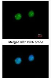
- Immunofluorescent analysis of Fibrillarin in paraformaldehyde-fixed HeLa cells using a Fibrillarin polyclonal antibody (Product # PA5-29801) at a 1:200 dilution.
Supportive validation
- Submitted by
- Invitrogen Antibodies (provider)
- Main image
- Experimental details
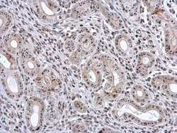
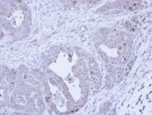
- Immunohistochemistry (Paraffin) analysis of Fibrillarin was performed in paraffin-embedded mouse cervix tissue using Fibrillarin Polyclonal Antibody (Product # PA5-29801) at a dilution of 1:500.
- Submitted by
- Invitrogen Antibodies (provider)
- Main image
- Experimental details
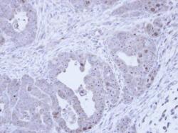
- Immunohistochemical analysis of paraffin-embedded NCIN87 Xenograft, using Fibrillarin (Product # PA5-29801) antibody at 1:100 dilution. Antigen Retrieval: Citrate buffer, pH 6.0, 15 min.
Supportive validation
- Submitted by
- Invitrogen Antibodies (provider)
- Main image
- Experimental details
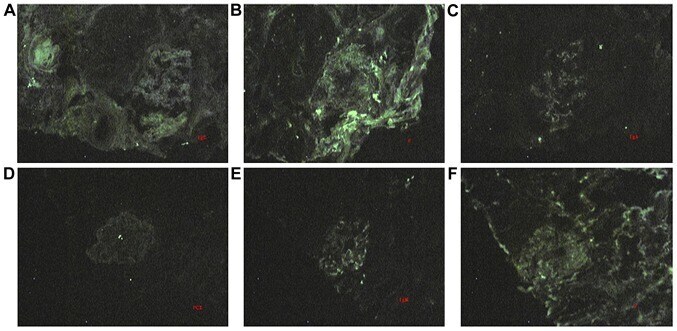
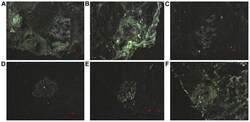
- Figure 5. Immunofluorescence staining of the kidney tissues in P17 and P18. (A) High deposition of IgG in the mesangial area. (B) Fib was deposited in the mesangial area as a mass. (C) IgA was deposited in the mesangial area as a mass. (D) No significant deposition of PCX. (E) High deposition of IgM in the mesangial zone. (F) High deposition of Fib in the mesangial zone. Magnification, x400. PCX, podocalyxin; Fib, fabrillarin.