 Explore
 Validate
 Learn
Learn Western blot
 ELISA
ELISAAntibody data
- Antibody Data
- Antigen structure
- References [0]
- Comments [0]
- Validations
- Western blot [3]
Submit
Validation data
Reference
Comment
Report error
- Product number
- PA5-22690 - Provider product page

- Provider
- Invitrogen Antibodies
- Product name
- ATM Polyclonal Antibody, DyLight™ 488
- Antibody type
- Polyclonal
- Antigen
- Other
- Reactivity
- Human, Mouse, Rat, Canine
- Host
- Rabbit
- Conjugate
- Green dye
- Isotype
- IgG
- Vial size
- 50 µL
- Concentration
- 1.47 mg/mL
- Storage
- 4° C, store in dark
No comments: Submit comment
Supportive validation
- Submitted by
- Invitrogen Antibodies (provider)
- Main image
- Experimental details
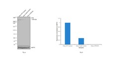
- Knockout of ATM was achieved by CRISPR-Cas9 genome editing using LentiArray™ Lentiviral sgRNA (Product # A32042, Assay ID CRISPR901163_LV) and LentiArray Cas9 Lentivirus (Product # A32064). Western blot analysis of ATM was performed by loading 30 µg of HeLa Wild Type (Lane 1), HeLa Cas9 (Lane 2) andHeLa ATM KO (Lane 3) whole cell extracts. The samples were electrophoresed using NuPAGE™ 3-8% Tris-Acetate Protein Gel (Product # EA0378BOX). Resolved proteins were then transferred onto a nitrocellulose membrane (Product # IB23001) by iBlot® 2 Dry Blotting System (Product # IB21001). The blot was probed with Anti-ATM Polyclonal Antibody, DyLight 488 (Product # PA5-22690, 1:1,000 dilution) and Goat anti-Rabbit IgG (H+L) Superclonal™ Recombinant Secondary Antibody, HRP (Product # A27036, 1:4,000 dilution) using the iBright FL 1000 (Product # A32752). Chemiluminescent detection was performed using Novex® ECL Chemiluminescent Substrate Reagent Kit (Product # WP20005). Loss of signal upon CRISPR mediated knockout (KO) using the LentiArray™ CRISPR product line confirms that antibody is specific to ATM.
- Conjugate
- Green dye

- Submitted by
- Invitrogen Antibodies (provider)
- Main image
- Experimental details
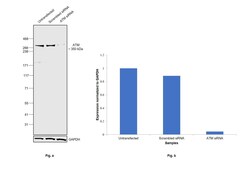
- Knockdown of Serine-protein kinase ATM was achieved by transfecting Hep G2 with Serine-protein kinase ATM specific siRNAs (Silencer® select Product # S1708, S1709). Western blot analysis (Fig. a) was performed using Whole cell extracts from the Serine-protein kinase ATM knockdown cells (lane 3), non-targeting scrambled siRNA transfected cells (lane 2) and untransfected cells (lane 1). The blot was probed with ATM Polyclonal Antibody, DyLight 488 (Product # PA5-22690, 1:1000 dilution ) and Goat anti-Rabbit IgG (H+L) Superclonal™ Recombinant Secondary Antibody, HRP (Product # A27036, 1:4000 dilution). Densitometric analysis of this western blot is shown in histogram (Fig. b). A decrease in signal upon siRNA mediated knockdown confirms that antibody is specific to Serine-protein kinase ATM.
- Conjugate
- Green dye

- Submitted by
- Invitrogen Antibodies (provider)
- Main image

- Experimental details
- Western blot was performed using Anti-ATM Polyclonal Antibody, DyLight 488 (Product # PA5-22690) and a 350kDa band corresponding to Serine-protein kinase ATM was observed across cell lines tested. Whole cell extracts (30 µg lysate) of Hep G2 (Lane 1), U-2 OS (Lane 2), HeLa (Lane 3), K-562 (Lane 4) and Jurkat (Lane 5) were electrophoresed using NuPAGE™ 3-8% Tris-Acetate Protein Gel (Product # EA0378BOX). Resolved proteins were then transferred onto a Nitrocellulose membrane (Product # IB23001) by iBlot® 2 Dry Blotting System (Product # IB21001). The blot was probed with the primary antibody (1:1000 dilution) and detected by chemiluminescence with Goat anti-Rabbit IgG (H+L) Superclonal™ Recombinant Secondary Antibody, HRP (Product # A27036, 1:4000 dilution) using the iBright FL 1000 (Product # A32752). Chemiluminescent detection was performed using Novex® ECL Reagent Kit (Product # WP20005).
- Conjugate
- Green dye
