 Explore
 Validate
 Learn
Learn Western blot
 Immunocytochemistry
 Immunoprecipitation
Antibody data
- Antibody Data
- Antigen structure
- References [4]
- Comments [0]
- Validations
- Immunocytochemistry [8]
- Other assay [1]
Submit
Validation data
Reference
Comment
Report error
- Product number
- MA1-2020 - Provider product page

- Provider
- Invitrogen Antibodies
- Product name
- Phospho-ATM (Ser1981) Monoclonal Antibody (10H11)
- Antibody type
- Monoclonal
- Antigen
- Synthetic peptide
- Description
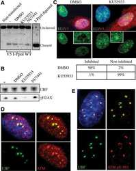
- MA1-2020 detects the phospho-ATM kinase in human and mouse samples. MA1-2020 has been successfully used in immunofluorescence, immunoprecipitation and Western blot procedures. By Western blot this antibody detects a ~370 kDa protein representing the phospho-ATM kinase in crude lysates from gamma irradiated HeLa cells. In immunofluorescence procedures, MA1-2020 recognizes the phospho-ATM kinase in irradiated human and mouse fibroblasts. The MA1-2020 immunogen is a phosphorylated synthetic peptide corresponding to the residues S(1974) L A F E E S(p) Q S T T I S S(1988) of human ATM Kinase protein.
- Reactivity
- Human, Mouse
- Host
- Mouse
- Isotype
- IgG
- Antibody clone number
- 10H11
- Vial size
- 200 μg
- Concentration
- 1 mg/mL
- Storage
- -20°C, Avoid Freeze/Thaw Cycles
Submitted references PARP1 promote autophagy in cardiomyocytes via modulating FoxO3a transcription.
A localized nucleolar DNA damage response facilitates recruitment of the homology-directed repair machinery independent of cell cycle stage.
2-Hydroxyethyl methacrylate-induced apoptosis through the ATM- and p53-dependent intrinsic mitochondrial pathway.
Disappearance of the telomere dysfunction-induced stress response in fully senescent cells.
Wang C, Xu W, Zhang Y, Zhang F, Huang K
Cell death & disease 2018 Oct 15;9(11):1047
Cell death & disease 2018 Oct 15;9(11):1047
A localized nucleolar DNA damage response facilitates recruitment of the homology-directed repair machinery independent of cell cycle stage.
van Sluis M, McStay B
Genes & development 2015 Jun 1;29(11):1151-63
Genes & development 2015 Jun 1;29(11):1151-63
2-Hydroxyethyl methacrylate-induced apoptosis through the ATM- and p53-dependent intrinsic mitochondrial pathway.
Schweikl H, Petzel C, Bolay C, Hiller KA, Buchalla W, Krifka S
Biomaterials 2014 Mar;35(9):2890-904
Biomaterials 2014 Mar;35(9):2890-904
Disappearance of the telomere dysfunction-induced stress response in fully senescent cells.
Bakkenist CJ, Drissi R, Wu J, Kastan MB, Dome JS
Cancer research 2004 Jun 1;64(11):3748-52
Cancer research 2004 Jun 1;64(11):3748-52
No comments: Submit comment
Supportive validation
- Submitted by
- Invitrogen Antibodies (provider)
- Main image

- Experimental details
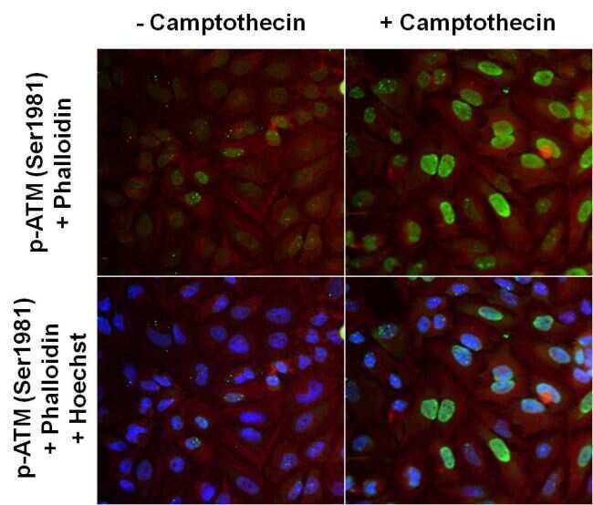
- Immunofluorescent analysis of ATM phosphorylated on pSer1981 (green) in HeLa cells left untreated (left panels) or treated with 10uM Camptothecin for 4 hours (right panels). Formalin fixed cells were permeabilized with 0.1% Triton X-100 in TBS for 10 minutes at room temperature and blocked with 1% Blocker BSA (Product # 37525) for 15 minutes at room temperature. Cells were probed with a phospho-ATM (pSer1981) monoclonal antibody (Product # MA1-2020), at a dilution of 1:1000 for at least 1 hour at room temperature, washed with PBS, and incubated with DyLight 488 goat anti-mouse IgG secondary antibody (Product # 35502) at a dilution of 1:400 for 30 minutes at room temperature. F-Actin (red) was stained with DyLight 554 Phalloidin (Product # 21834) (top panels). A merged image with nuclei staining (blue) using Hoechst 33342 dye (Product # 62249) is shown on bottom panels. Images were taken on a Thermo Scientific ArrayScan at 20X magnification.
- Submitted by
- Invitrogen Antibodies (provider)
- Main image

- Experimental details
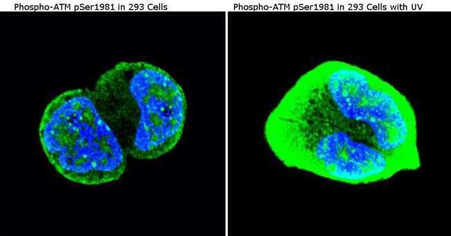
- Immunofluorescent analysis of Phospho-ATM pSer1981 in 293 untreated cells (left panel) or stimulated cells with 100 mJ UV (right panel). Formalin-fixed cells were permeabilized with 0.1% Triton X-100 in TBS for 5-10 minutes at room temperature and blocked with 3% BSA-PBS for 30 minutes at room temperature. Cells were probed with a Phospho-ATM pSer1981 Monclonal Antibody (10H11) (Product # MA1-2020) at a dilution of 1:50 and incubated overnight in a humidified chamber. Cells were washed with PBST and incubated with a DyLight-conjugated secondary antibody for 45 minutes at room temperature in the dark. F-actin (red) was stained with a fluorescent phalloidin and nuclei (blue) were stained with DAPI. Images were taken at a 60X magnification.
- Submitted by
- Invitrogen Antibodies (provider)
- Main image
- Experimental details
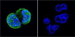
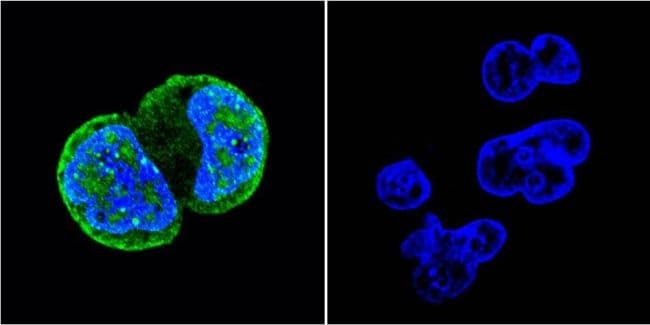
- Immunofluorescent analysis of Phospho-ATM pSer1981 (green) showing staining in HEK-293 cells (right) compared to a negative control without primary antibody (left). Formalin-fixed cells were permeabilized with 0.1% Triton X-100 in TBS for 5-10 minutes and blocked with 3% BSA-PBS for 30 minutes at room temperature. Cells were probed with a Phospho-ATM pSer1981 monoclonal antibody (Product # MA1-2020) in 3% BSA-PBS at a dilution of 1:50 and incubated overnight at 4°C in a humidified chamber. Cells were washed with PBST and incubated with a DyLight-conjugated secondary antibody in PBS at room temperature in the dark. F-actin (red) was stained with a fluorescent red phalloidin and nuclei (blue) were stained with Hoechst or DAPI. Images were taken at a magnification of 60x.
- Submitted by
- Invitrogen Antibodies (provider)
- Main image
- Experimental details
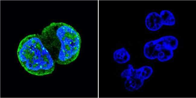
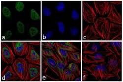
- Immunofluorescence analysis of Phospho-ATM pSer1981 was performed using 70% confluent log phase HeLa cells irradiated with UV for 4 hours. The cells were fixed with 4% paraformaldehyde for 10 minutes, permeabilized with 0.1% Triton™ X-100 for 10 minutes, and blocked with 1% BSA for 1 hour at room temperature. The cells were labeled with Phospho-ATM (Ser 1981) (10H11) Mouse Monoclonal Antibody (Product # MA1-2020) at 1:250 dilution in 0.1% BSA and incubated for 3 hours at room temperature and then labeled with Goat anti-Mouse IgG (H+L) Superclonal™ Secondary Antibody, Alexa Fluor® 488 conjugate (Product # A28175) at a dilution of 1:2000 for 45 minutes at room temperature (Panel a: green). Nuclei (Panel b: blue) were stained with SlowFade® Gold Antifade Mountant with DAPI (Product # S36938). F-actin (Panel c: red) was stained with Rhodamine Phalloidin (Product # R415, 1:300). Panel d represents the merged image showing nuclear localization. Panel e shows the untreated cells. Panel f represents the no primary antibody control. The images were captured at 60X magnification.
- Submitted by
- Invitrogen Antibodies (provider)
- Main image

- Experimental details
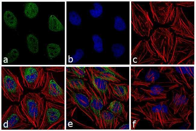
- Immunofluorescent analysis of Phospho-ATM pSer1981 (green) showing staining in HEK-293 cells (right) compared to a negative control without primary antibody (left). Formalin-fixed cells were permeabilized with 0.1% Triton X-100 in TBS for 5-10 minutes and blocked with 3% BSA-PBS for 30 minutes at room temperature. Cells were probed with a Phospho-ATM pSer1981 monoclonal antibody (Product # MA1-2020) in 3% BSA-PBS at a dilution of 1:50 and incubated overnight at 4°C in a humidified chamber. Cells were washed with PBST and incubated with a DyLight-conjugated secondary antibody in PBS at room temperature in the dark. F-actin (red) was stained with a fluorescent red phalloidin and nuclei (blue) were stained with Hoechst or DAPI. Images were taken at a magnification of 60x.
- Submitted by
- Invitrogen Antibodies (provider)
- Main image

- Experimental details
- Immunofluorescent analysis of Phospho-ATM pSer1981 in 293 untreated cells (left panel) or stimulated cells with 100 mJ UV (right panel). Formalin-fixed cells were permeabilized with 0.1% Triton X-100 in TBS for 5-10 minutes at room temperature and blocked with 3% BSA-PBS for 30 minutes at room temperature. Cells were probed with a Phospho-ATM pSer1981 Monclonal Antibody (10H11) (Product # MA1-2020) at a dilution of 1:50 and incubated overnight in a humidified chamber. Cells were washed with PBST and incubated with a DyLight-conjugated secondary antibody for 45 minutes at room temperature in the dark. F-actin (red) was stained with a fluorescent phalloidin and nuclei (blue) were stained with DAPI. Images were taken at a 60X magnification.

- Submitted by
- Invitrogen Antibodies (provider)
- Main image

- Experimental details
- Immunofluorescent analysis of ATM phosphorylated on pSer1981 (green) in HeLa cells left untreated (left panels) or treated with 10uM Camptothecin for 4 hours (right panels). Formalin fixed cells were permeabilized with 0.1% Triton X-100 in TBS for 10 minutes at room temperature and blocked with 1% Blocker BSA (Product # 37525) for 15 minutes at room temperature. Cells were probed with a phospho-ATM (pSer1981) monoclonal antibody (Product # MA1-2020), at a dilution of 1:1000 for at least 1 hour at room temperature, washed with PBS, and incubated with DyLight 488 goat anti-mouse IgG secondary antibody (Product # 35502) at a dilution of 1:400 for 30 minutes at room temperature. F-Actin (red) was stained with DyLight 554 Phalloidin (Product # 21834) (top panels). A merged image with nuclei staining (blue) using Hoechst 33342 dye (Product # 62249) is shown on bottom panels. Images were taken on a Thermo Scientific ArrayScan at 20X magnification.

Supportive validation
- Submitted by
- Invitrogen Antibodies (provider)
- Main image
- Experimental details
- NULL
