Explore
 Validate
 Learn
Learn Western blot
 Other assay
Antibody data
- Antibody Data
- Antigen structure
- References [1]
- Comments [0]
- Validations
- Other assay [1]
Submit
Validation data
Reference
Comment
Report error
- Product number
- MA5-14871 - Provider product page

- Provider
- Invitrogen Antibodies
- Product name
- ATM Monoclonal Antibody (C.74.4)
- Antibody type
- Monoclonal
- Antigen
- Recombinant full-length protein
- Description
- It is not recommended to aliquot this antibody.
- Reactivity
- Human, Mouse
- Host
- Rabbit
- Isotype
- IgG
- Antibody clone number
- C.74.4
- Vial size
- 100 μL
- Concentration
- 143 μg/mL
- Storage
- -20°C
Submitted references Phosphorylation of BRCA1 by ATM upon double-strand breaks impacts ATM function in end-resection: A potential feedback loop.
Qi L, Chakravarthy R, Li MM, Deng CX, Li R, Hu Y
iScience 2022 Sep 16;25(9):104944
iScience 2022 Sep 16;25(9):104944
No comments: Submit comment
Supportive validation
- Submitted by
- Invitrogen Antibodies (provider)
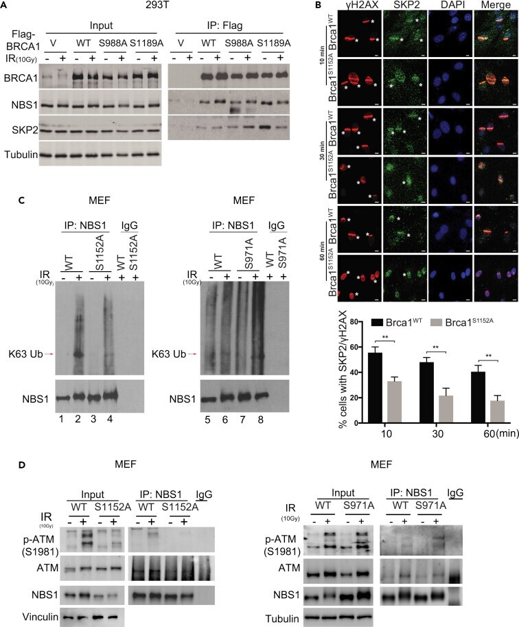
- Main image

- Experimental details
- Abrogation of ATM phosphorylation on BRCA1-S1152 reduces SKP2 recruitment and ATM-NBS1 interaction upon DNA damage (A) Coimmunoprecipitation shows reduced interaction between BRCA1-S1198A and SKP2 in HEK293 cells. (B) Recruitment of SKP2 upon microirradiation. SKP2 laser stripes (green) and gammaH2AX (red) at 10, 30, and 60 min time points are shown. Scale bar, 10 mum. Bar graph below is quantification of three independent experiments. For 10 min (n = 326 for Brca1 WT and n = 351 for Brca1 S1152A/S1152A , p = 0.007656); 30 min (n = 336 for Brca1 WT and n = 427 for Brca1 S1152A/S1152A , p = 0.009519); 60 min (n = 336 for Brca1 WT and n = 380 for Brca1 S1152A/S1152A , p = 0.009258). (C) Immunoprecipitation of endogenous NBS1 shows reduced K63-linked ubiquitination of NBS1 in Brca1 S1152A/S1152A MEF cells, but not in Brca1 S971A/S971A MEF cells, in response to DNA damage. (D) Coimmunoprecipitation shows reduced interaction between NBS1 and ATM upon DNA damage in Brca1 S1152A/S1152A MEF cells but not in Brca1 S971A/S971A MEF cells.