Explore
 Validate
 Learn
Learn Western blot
Antibody data
- Antibody Data
- Antigen structure
- References [1]
- Comments [0]
- Validations
- Western blot [4]
- Immunocytochemistry [1]
- Immunohistochemistry [6]
Submit
Validation data
Reference
Comment
Report error
- Product number
- GTX100605 - Provider product page

- Provider
- GeneTex
- Proper citation
- GeneTex Cat#GTX100605, RRID:AB_1241140
- Product name
- p130Cas antibody [N2C2], Internal
- Antibody type
- Polyclonal
- Reactivity
- Human, Mouse, Rat
- Host
- Rabbit
Submitted references N-WASP-directed actin polymerization activates Cas phosphorylation and lamellipodium spreading.
Zhang X, Moore SW, Iskratsch T, Sheetz MP
Journal of cell science 2014 Apr 1;127(Pt 7):1394-405
Journal of cell science 2014 Apr 1;127(Pt 7):1394-405
No comments: Submit comment
Supportive validation
- Submitted by
- GeneTex (provider)
- Main image

- Experimental details
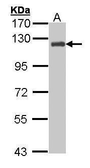
- Sample (30 ug of whole cell lysate) A: H1299 7.5% SDS PAGE GTX100605 diluted at 1:1000
- Submitted by
- GeneTex (provider)
- Main image
- Experimental details
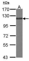
- Sample (30 ug of whole cell lysate) A:NIH-3T37.5% SDS PAGE GTX100605 diluted at 1:1000

- Submitted by
- GeneTex (provider)
- Main image
- Experimental details
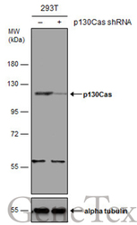
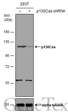
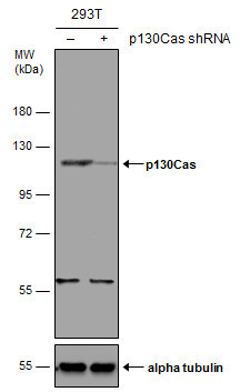
- Non-transfected (¡V) and transfected (+) 293T whole cell extracts (30 ?g) were separated by 7.5% SDS-PAGE, and the membrane was blotted with p130Cas antibody [N2C2], Internal (GTX100605) diluted at 1:500. The HRP-conjugated anti-rabbit IgG antibody (GTX213110-01) was used to detect the primary antibody.
- Submitted by
- GeneTex (provider)
- Main image
- Experimental details
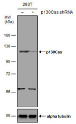
- Non-transfected (¡V) and transfected (+) 293T whole cell extracts (30 ?g) were separated by 7.5% SDS-PAGE, and the membrane was blotted with p130Cas antibody [N2C2], Internal (GTX100605) diluted at 1:500. The HRP-conjugated anti-rabbit IgG antibody (GTX213110-01) was used to detect the primary antibody.
Supportive validation
- Submitted by
- GeneTex (provider)
- Main image
- Experimental details
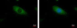
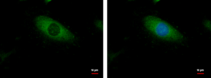
- BCAR1 antibody [N2C2], Internal detects BCAR1 protein at cytoplasm by immunofluorescent analysis.Sample: HeLa cells were fixed in ice-cold MeOH for 5 min.Green: BCAR1 protein stained by BCAR1 antibody [N2C2], Internal (GTX100605) diluted at 1:500.Blue: Hoechst 33342 staining.
Supportive validation
- Submitted by
- GeneTex (provider)
- Main image

- Experimental details
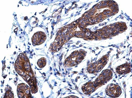
- BCAR1 antibody [N2C2], Internal detects BCAR1 protein at cytoplasm on human breast carcinoma by immunohistochemical analysis. Sample: Paraffin-embedded human breast carcinoma. BCAR1 antibody [N2C2], Internal (GTX100605) diluted at 1:500.
- Submitted by
- GeneTex (provider)
- Main image
- Experimental details
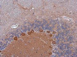
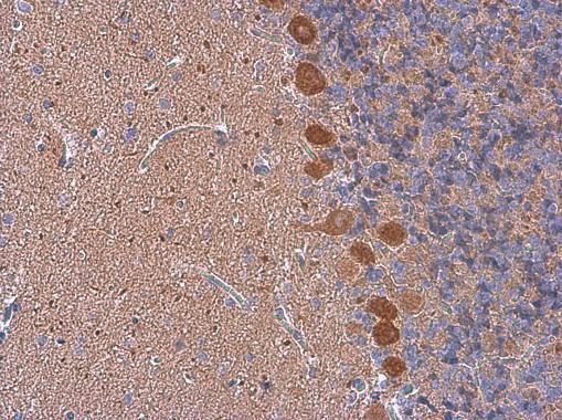
- p130Cas antibody [N2C2], Internal detects p130Cas protein at cytoplasm in mouse brain by immunohistochemical analysis. Sample: Paraffin-embedded mouse brain. p130Cas antibody [N2C2], Internal (GTX100605) diluted at 1:500.
- Submitted by
- GeneTex (provider)
- Main image

- Experimental details
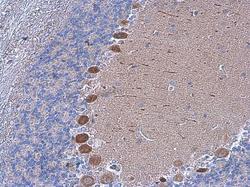
- p130Cas antibody [N2C2], Internal detects p130Cas protein at cytoplasm in rat brain by immunohistochemical analysis. Sample: Paraffin-embedded rat brain. p130Cas antibody [N2C2], Internal (GTX100605) diluted at 1:500.
- Submitted by
- GeneTex (provider)
- Main image
- Experimental details
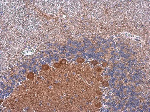
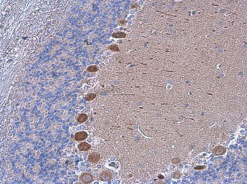
- p130Cas antibody [N2C2], Internal detects p130Cas protein at cytoplasm in mouse brain by immunohistochemical analysis. Sample: Paraffin-embedded mouse brain. p130Cas antibody [N2C2], Internal (GTX100605) diluted at 1:500.
- Submitted by
- GeneTex (provider)
- Main image

- Experimental details
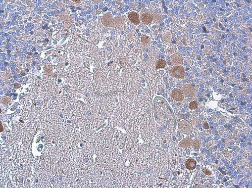
- p130Cas antibody [N2C2], Internal detects p130Cas protein at cytoplasm in rat brain by immunohistochemical analysis. Sample: Paraffin-embedded rat brain. p130Cas antibody [N2C2], Internal (GTX100605) diluted at 1:500.
- Submitted by
- GeneTex (provider)
- Main image
- Experimental details
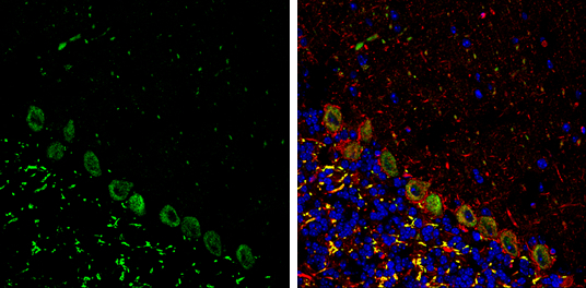
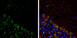
- p130Cas antibody [N2C2], Internal detects p130Cas protein expression by immunohistochemical analysis.Sample: Frozen-sectioned adult mouse cerebellum. Green: p130Cas protein stained by p130Cas antibody [N2C2], Internal (GTX100605) diluted at 1:250.Red: NF-H, stained by NF-H antibody [GT114] (GTX634289) diluted at 1:500.Blue: Fluoroshield with DAPI (GTX30920).