Explore
 Validate
 Learn
LearnMA5-17093
antibody from Invitrogen Antibodies
Targeting: CDKN2A
ARF, CDK4I, CDKN2, CMM2, INK4, INK4a, MLM, MTS1, p14, p14ARF, p16, p16INK4a, p19, p19Arf
 Western blot
Antibody data
- Antibody Data
- Antigen structure
- References [5]
- Comments [0]
- Validations
- Western blot [1]
- Immunohistochemistry [2]
- Other assay [3]
Submit
Validation data
Reference
Comment
Report error
- Product number
- MA5-17093 - Provider product page

- Provider
- Invitrogen Antibodies
- Product name
- p16INK4a Monoclonal Antibody (5A8A4)
- Antibody type
- Monoclonal
- Antigen
- Purifed from natural sources
- Description
- MA5-17093 targets P16 in IHC and WB applications and shows reactivity with Human samples.
- Antibody clone number
- 5A8A4
- Concentration
- 1.0 mg/mL
Submitted references Neutrophils infiltration and its correlation with human papillomavirus status in the oral squamous cell carcinoma.
Epithelial innate immunity mediates tubular cell senescence after kidney injury.
Defining UHRF1 Domains that Support Maintenance of Human Colon Cancer DNA Methylation and Oncogenic Properties.
Reduced levels of methyltransferase DNMT2 sensitize human fibroblasts to oxidative stress and DNA damage that is accompanied by changes in proliferation-related miRNA expression.
Metastatic model of HPV+ oropharyngeal squamous cell carcinoma demonstrates heterogeneity in tumor metastasis.
Li C, Zhao L, Wang Q, Ma S, Sun J, Ma C, Liu J, Jing X, Ai D, Nan Z, Qu X
Cancer management and research 2019;11:5171-5185
Cancer management and research 2019;11:5171-5185
Epithelial innate immunity mediates tubular cell senescence after kidney injury.
Jin H, Zhang Y, Ding Q, Wang SS, Rastogi P, Dai DF, Lu D, Purvis M, Cao C, Wang A, Liu D, Ren C, Elhadi S, Hu MC, Chai Y, Zepeda-Orozco D, Campisi J, Attanasio M
JCI insight 2019 Jan 24;4(2)
JCI insight 2019 Jan 24;4(2)
Defining UHRF1 Domains that Support Maintenance of Human Colon Cancer DNA Methylation and Oncogenic Properties.
Kong X, Chen J, Xie W, Brown SM, Cai Y, Wu K, Fan D, Nie Y, Yegnasubramanian S, Tiedemann RL, Tao Y, Chiu Yen RW, Topper MJ, Zahnow CA, Easwaran H, Rothbart SB, Xia L, Baylin SB
Cancer cell 2019 Apr 15;35(4):633-648.e7
Cancer cell 2019 Apr 15;35(4):633-648.e7
Reduced levels of methyltransferase DNMT2 sensitize human fibroblasts to oxidative stress and DNA damage that is accompanied by changes in proliferation-related miRNA expression.
Lewinska A, Adamczyk-Grochala J, Kwasniewicz E, Deregowska A, Semik E, Zabek T, Wnuk M
Redox biology 2018 Apr;14:20-34
Redox biology 2018 Apr;14:20-34
Metastatic model of HPV+ oropharyngeal squamous cell carcinoma demonstrates heterogeneity in tumor metastasis.
Vermeer DW, Coppock JD, Zeng E, Lee KM, Spanos WC, Onken MD, Uppaluri R, Lee JH, Vermeer PD
Oncotarget 2016 Apr 26;7(17):24194-207
Oncotarget 2016 Apr 26;7(17):24194-207
No comments: Submit comment
Supportive validation
- Submitted by
- Invitrogen Antibodies (provider)
- Main image
- Experimental details
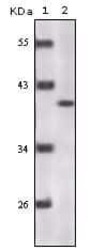
- Western blot analysis of P16 using a P16 monoclonal antibody (Product # MA5-17093) against a truncated P16 recombinant protein.

Supportive validation
- Submitted by
- Invitrogen Antibodies (provider)
- Main image
- Experimental details
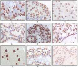
- Immunohistochemical analysis of paraffin-embedded human lung adenocarcinoma (A), esophageal squamous cell carcinoma (B), hepatic cell carcinoma (C), thyroid tumor (D), breast adenofibroma (E), breast infiltrating ductal carcinoma (F), normal cerebrum tissue (G), normal colon tissue (H), normal esophageal tissue (I) using P16 monoclonal antibody (Product # MA5-17093) followed with DAB staining.
- Submitted by
- Invitrogen Antibodies (provider)
- Main image
- Experimental details
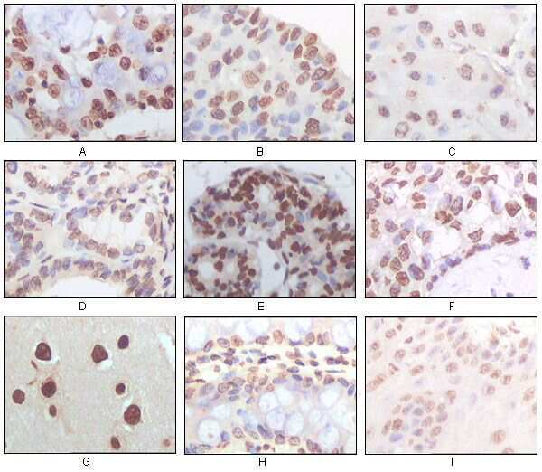
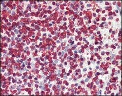
- Immunohistochemical analysis of paraffin-embedded human spleen tissues using P16 monoclonal antibody (Product # MA5-17093).

Supportive validation
- Submitted by
- Invitrogen Antibodies (provider)
- Main image

- Experimental details
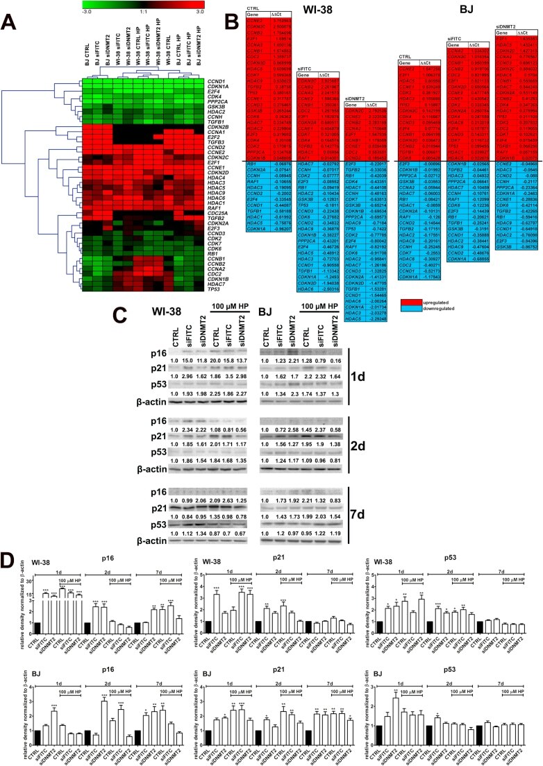
- Fig. 3 DNMT2 silencing-mediated effects on mRNA (A, B) and protein levels (C, D) of cell cycle regulators. HP treatment is also considered. (A, B) The expression of selected genes involved in the regulation of cell cycle. Cells were treated with 100 muM HP for 2 h and then cultured for 24 h. (A) A heat map generated from qRT-PCR data is shown. Hierarchical clustering was created using Genesis 1.7.7 software. (B) HP-mediated up- (red) and downregulation (blue) of cell cycle genes. DeltaDeltaCt values are shown. (C, D) Western blot analysis of the levels of p16, p21 and p53. Data were normalized to beta-actin. Bars indicate SD, n = 3, * p < 0.05, ** p < 0.01, *** p < 0.001 compared to CTRL (ANOVA and Dunnett's a posteriori test). Proliferatively active fibroblasts were used, namely WI-38 cells at PDLs from 34 to 43 and BJ cells at PDLs from 27 to 43. CTRL, non-transfected control fibroblasts; siFITC, fibroblasts transfected with control FITC-conjugated siRNA; siDNMT2, fibroblasts transfected with DNMT2 siRNA. (For interpretation of the references to color in this figure legend, the reader is referred to the web version of this article.) Fig. 3
- Submitted by
- Invitrogen Antibodies (provider)
- Main image

- Experimental details
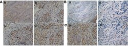
- Figure 3 Protein expression in mEERL, MLMs and human HPV+ OPSCC ( A ) Heat map of reverse phase protein array analysis of parental mEERL cells and MLM clones performed in triplicate. ( B ) Immunohistochemical staining of normal tissue (The Human Protein Atlas) (38), human HPV+ OPSCC and mEERL tumor for hallmark proteins of OPSCC (Keratin, E-cadherin, P16, BRCA2 and EGFR). Scale bar, 40 mum. ( C ) RPPA heatmap of protein expression of four markers (EGFR, BRCA2, E-cadherin and P16) analyzed in panel B. Cytokeratin was not analyzed as it was not included in the RPPA.

- Submitted by
- Invitrogen Antibodies (provider)
- Main image
- Experimental details
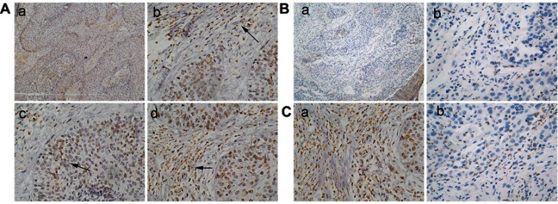
- Figure 1 Immunohistochemistry staining results of CD15 and HPV. ( A ) There were many CD15+ neutrophils (black arrow) in OSCC tissues ( a ). In tumors areas, some neutrophils lied in the stroma of tumors ( b ), some were located within the carcinoma nests ( c ), and some infiltrated in the borderline of tumor invasion ( d ). a: 100x; b, c, d: 400x. ( B ) The expression of HPV in peritumoral tissues was negative or weak ( a ). In OSCC tissues, HPV expression was obviously strong and located mainly in the cytoplasm of tumor cells ( b ). a: 100x; b: 400x. ( C ) The relationship of HPV expression on tumor cells and infiltration of neutrophils. In HPV-negative expression tumors, there were more neutrophils ( a ); while in HPV-positive expression tumors, there were relatively fewer neutrophils ( b ). a, b: 400x. Abbreviations: OSCC, oral squamous cell carcinoma; HPV, Human papillomavirus.