 Explore
 Validate
 Learn
LearnMA5-17142
antibody from Invitrogen Antibodies
Targeting: CDKN2A
ARF, CDK4I, CDKN2, CMM2, INK4, INK4a, MLM, MTS1, p14, p14ARF, p16, p16INK4a, p19, p19Arf
 Western blot
Antibody data
- Antibody Data
- Antigen structure
- References [4]
- Comments [0]
- Validations
- Western blot [1]
- Immunohistochemistry [1]
- Other assay [3]
Submit
Validation data
Reference
Comment
Report error
- Product number
- MA5-17142 - Provider product page

- Provider
- Invitrogen Antibodies
- Product name
- p16INK4a Monoclonal Antibody (1E12E10)
- Antibody type
- Monoclonal
- Antigen
- Purifed from natural sources
- Description
- MA5-17142 targets P16 in IHC and WB applications and shows reactivity with Human, Mouse, and Rat samples.
- Antibody clone number
- 1E12E10
- Concentration
- Conc. Not Determined
Submitted references Hypoxia-inducible factor-2α mediates senescence-associated intrinsic mechanisms of age-related bone loss.
Cdkn2a Loss in a Model of Neurofibroma Demonstrates Stepwise Tumor Progression to Atypical Neurofibroma and MPNST.
TGF-β1/IL-11/MEK/ERK signaling mediates senescence-associated pulmonary fibrosis in a stress-induced premature senescence model of Bmi-1 deficiency.
Chronic Intermittent Hypoxia Triggers a Senescence-like Phenotype in Human White Preadipocytes.
Lee SY, Park KH, Lee G, Kim SJ, Song WH, Kwon SH, Koh JT, Huh YH, Ryu JH
Experimental & molecular medicine 2021 Apr;53(4):591-604
Experimental & molecular medicine 2021 Apr;53(4):591-604
Cdkn2a Loss in a Model of Neurofibroma Demonstrates Stepwise Tumor Progression to Atypical Neurofibroma and MPNST.
Chaney KE, Perrino MR, Kershner LJ, Patel AV, Wu J, Choi K, Rizvi TA, Dombi E, Szabo S, Largaespada DA, Ratner N
Cancer research 2020 Nov 1;80(21):4720-4730
Cancer research 2020 Nov 1;80(21):4720-4730
TGF-β1/IL-11/MEK/ERK signaling mediates senescence-associated pulmonary fibrosis in a stress-induced premature senescence model of Bmi-1 deficiency.
Chen H, Chen H, Liang J, Gu X, Zhou J, Xie C, Lv X, Wang R, Li Q, Mao Z, Sun H, Zuo G, Miao D, Jin J
Experimental & molecular medicine 2020 Jan;52(1):130-151
Experimental & molecular medicine 2020 Jan;52(1):130-151
Chronic Intermittent Hypoxia Triggers a Senescence-like Phenotype in Human White Preadipocytes.
Polonis K, Becari C, Chahal CAA, Zhang Y, Allen AM, Kellogg TA, Somers VK, Singh P
Scientific reports 2020 Apr 22;10(1):6846
Scientific reports 2020 Apr 22;10(1):6846
No comments: Submit comment
Supportive validation
- Submitted by
- Invitrogen Antibodies (provider)
- Main image

- Experimental details
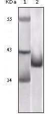
- Western blot analysis of P16 using a P16 monoclonal antibody (Product # MA5-17142) against a GST-tagged truncated P16 recombinant protein.
Supportive validation
- Submitted by
- Invitrogen Antibodies (provider)
- Main image
- Experimental details
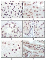
- Immunohistochemical analysis of paraffin-embedded rat liver tissue (A), human brain tumor (B), breast cancer (C), esophageal epithelium tissue (D), mouse brain tissue (E) and stomach tisue (F) using P16 monoclonal antibody (Product # MA5-17142) followed with DAB staining.

Supportive validation
- Submitted by
- Invitrogen Antibodies (provider)
- Main image
- Experimental details
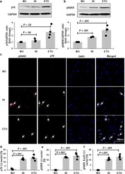
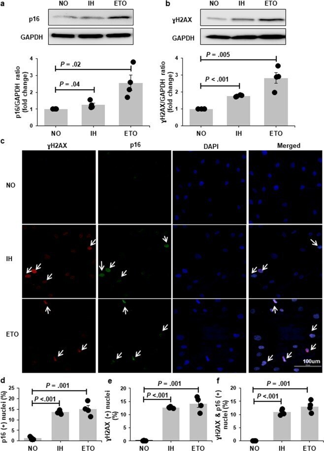
- Figure 2 P16 and gammaH2AX expression is increased with chronic exposure to intermittent hypoxia (IH). Representative Western bolts and graphs showing upregulation of p16 ( a ) and gammaH2AX ( b ) protein expression with IH treatment. Representative confocal images showing increased nuclear localization of p16 (green) and gammaH2AX (red) in cells exposed to IH ( c ). Nuclei are counterstained blue (DAPI). White arrows indicate positive nuclei. Quantitation of cells positive for nuclear p16 ( d ), gammaH2AX ( e ) and p16&gammaH2AX ( f ) in preadipocytes grown in continuous normoxia (NO) versus cells grown with intermittent exposure to hypoxia (n = 4 independent experiments). Cells treated with etopside (ETO) were used as positive control. Data are presented as mean +- SEM. P -values determined by one-tailed paired t-test compared to the NO control.
- Submitted by
- Invitrogen Antibodies (provider)
- Main image

- Experimental details
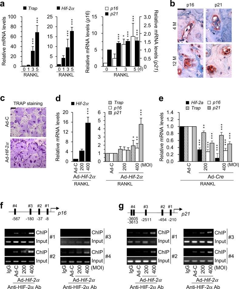
- Fig. 5 Upregulation of HIF-2alpha expression by aging stimulates osteoclastogenesis and osteoclast senescence. a The mRNA levels of osteoclast marker genes ( Trap ), Hif-2 alpha, and senescence markers ( p16 , p21 ) during M-CSF/RANKL-induced osteoclastogenesis of bone marrow macrophages (BMMs) ( n >= 3). b Representative images of p16 and p21 immunostaining in bone osteoclasts from young (4-month-old) and old (12-month-old) mice. Scale bar: 25 mum. M month. c, d TRAP staining ( c ) and qRT-PCR analysis of Hif-2alpha , Trap , p16 , and p21 expression ( n >= 4; d ) in Hif-2 alpha-overexpressing osteoclasts. BMMs were infected with Ad-C or Ad- Hif-2alpha and cultured with M-CSF and RANKL for 5 days. Scale bar: 100 mum. e The mRNA levels of Hif-2alpha , Trap, p16 , and p21 . Osteoclasts isolated from Hif-2alpha fl/fl mice were infected with Ad-C or Ad- Cre during osteoclastogenesis ( n >= 4). f, g Binding of HIF-2alpha to the promoter regions of p16 ( f ) and p21 ( g ). Ad- Hif-2alpha -infected osteoclasts were subjected to ChIP with an anti-HIF-2alpha antibody and a primer pair designed to span the putative HIF-2alpha binding regions [5''-(A/G)CGTG-3''] within the promoters of p16 ( n = 3; f ) and p21 ( n = 3; g ). The values are presented as the means +- SDs (* P < 0.05, ** P < 0.01, and *** P < 0.005).
- Submitted by
- Invitrogen Antibodies (provider)
- Main image
- Experimental details
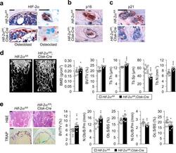
- Fig. 6 Osteoclast-specific depletion of HIF-2alpha increases bone mass in aged mice. a - c Immunostaining of HIF-2alpha, p16, and p21 in Hif-2alpha fl/fl and Hif-2alpha ; fl/fl Ctsk - Cre mice. Osteoclast-specific depletion of HIF-2alpha in 12-month-old Hif-2alpha fl/fl and Hif-2alpha ; fl/fl Ctsk - Cre mice was assessed by immunohistochemistry with anti-HIF-2alpha antibody. The dotted lines indicate osteoblasts (Scale bar: 25 mum; a ). Immunostaining of p16 ( b ) and p21 ( c ) in osteoclasts from Hif-2alpha fl/fl and Hif-2alpha ; fl/fl Ctsk - Cre mice was examined by immunofluorescence microscopy. Scale bar: 25 mum. d Quantitative uCT analysis of trabecular bones. BMD, BV/TV, Tb.Th, Tb.Sp, and Tb.N in trabecular bones from 12-month-old Hif-2alpha fl/fl and Hif-2alpha ; fl/fl Ctsk-Cre mice ( n = 8). e Representative images of H&E and TRAP staining in 12-month-old Hif-2alpha fl/fl and Hif-2alpha ; fl/fl Ctsk-Cre mice ( n = 8; Scale bar, 100 mum). BV/TV, N.Ob/B.Pm, Ob.S/BS, N.Oc/B.Pm, and Oc.S/BS were determined by bone histomorphometric analyses of the metaphyseal regions of femurs. The values are presented as the means +- SDs (* P < 0.05, ** P < 0.01, *** P < 0.005).
