Explore
 Validate
 Learn
LearnAF5779
antibody from R&D Systems
Targeting: CDKN2A
ARF, CDK4I, CDKN2, CMM2, INK4, INK4a, MLM, MTS1, p14, p14ARF, p16, p16INK4a, p19, p19Arf
 Western blot
Antibody data
- Antibody Data
- Antigen structure
- References [3]
- Comments [0]
- Validations
- Western blot [2]
- Immunocytochemistry [1]
Submit
Validation data
Reference
Comment
Report error
- Product number
- AF5779 - Provider product page

- Provider
- R&D Systems
- Product name
- Human p16INK4a/ CDKN2A Antibody
- Antibody type
- Polyclonal
- Description
- Antigen Affinity-purified. Detects human p16INK4a/CDKN2A in Western blots.
- Reactivity
- Human
- Host
- Goat
- Conjugate
- Unconjugated
- Antigen sequence
P42771- Isotype
- IgG
- Vial size
- 100 ug
- Concentration
- LYOPH
- Storage
- Use a manual defrost freezer and avoid repeated freeze-thaw cycles. 12 months from date of receipt, -20 to -70 °C as supplied. 1 month, 2 to 8 °C under sterile conditions after reconstitution. 6 months, -20 to -70 °C under sterile conditions after reconstitution.
Submitted references MAX inactivation is an early event in GIST development that regulates p16 and cell proliferation.
Adiponectin corrects premature cellular senescence and normalizes antimicrobial peptide levels in senescent keratinocytes.
Induction of heparanase by HPV E6 oncogene in head and neck squamous cell carcinoma.
Schaefer IM, Wang Y, Liang CW, Bahri N, Quattrone A, Doyle L, Mariño-Enríquez A, Lauria A, Zhu M, Debiec-Rychter M, Grunewald S, Hechtman JF, Dufresne A, Antonescu CR, Beadling C, Sicinska ET, van de Rijn M, Demetri GD, Ladanyi M, Corless CL, Heinrich MC, Raut CP, Bauer S, Fletcher JA
Nature communications 2017 Mar 8;8:14674
Nature communications 2017 Mar 8;8:14674
Adiponectin corrects premature cellular senescence and normalizes antimicrobial peptide levels in senescent keratinocytes.
Jin T, Kim MJ, Heo WI, Park KY, Choi SY, Lee MK, Hong SP, Kim SJ, Im M, Moon NJ, Seo SJ
Biochemical and biophysical research communications 2016 Sep 2;477(4):678-684
Biochemical and biophysical research communications 2016 Sep 2;477(4):678-684
Induction of heparanase by HPV E6 oncogene in head and neck squamous cell carcinoma.
Hirshoren N, Bulvik R, Neuman T, Rubinstein AM, Meirovitz A, Elkin M
Journal of cellular and molecular medicine 2014 Jan;18(1):181-6
Journal of cellular and molecular medicine 2014 Jan;18(1):181-6
No comments: Submit comment
Supportive validation
- Submitted by
- R&D Systems (provider)
- Main image
- Experimental details
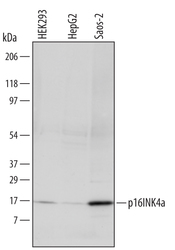
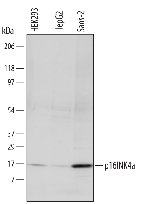
- Detection of Human p16INK4a/CDKN2A by Western Blot. Western blot shows lysates of HEK293 human embryonic kidney cell line, HepG2 human hepatocellular carcinoma cell line, and Saos-2 human osteosarcoma cell line. PVDF membrane was probed with 1 µg/mL of Goat Anti-Human p16INK4a/CDKN2A Antigen Affinity-purified Polyclonal Antibody (Catalog # AF5779) followed by HRP-conjugated Anti-Goat IgG Secondary Antibody (Catalog # HAF109). A specific band was detected for p16INK4a/CDKN2A at approximately 16 kDa (as indicated). This experiment was conducted under reducing conditions and using Immunoblot Buffer Group 1.
- Submitted by
- R&D Systems (provider)
- Main image
- Experimental details
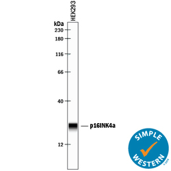
- Detection of Human p16INK4a/ CDKN2A by Simple WesternTM. Simple Western lane view shows lysates of HEK293 human embryonic kidney cell line, loaded at 0.2 mg/mL. A specific band was detected for p16INK4a/ CDKN2A at approximately 24 kDa (as indicated) using 10 µg/mL of Goat Anti-Human p16INK4a/ CDKN2A Antigen Affinity-purified Polyclonal Antibody (Catalog # AF5779) followed by 1:50 dilution of HRP-conjugated Anti-Goat IgG Secondary Antibody (Catalog # HAF109). This experiment was conducted under reducing conditions and using the 12-230 kDa separation system.

Supportive validation
- Submitted by
- R&D Systems (provider)
- Main image

- Experimental details
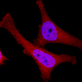
- p16INK4a / CDKN2A in HeLa Human Cell Line. p16INK4a / CDKN2A was detected in immersion fixed HeLa human cervical epithelial carcinoma cell line using Goat Anti-Human p16INK4a/ CDKN2A Antigen Affinity-purified Polyclonal Antibody (Catalog # AF5779) at 0.3 µg/mL for 3 hours at room temperature. Cells were stained using the NorthernLights™ 557-conjugated Anti-Goat IgG Secondary Antibody (red; Catalog # NL001) and counterstained with DAPI (blue). Specific staining was localized to cytoplasm and nuclei. View our protocol for Fluorescent ICC Staining of Cells on Coverslips.