 Explore
 Validate
 Learn
Learn Western blot
 Immunoprecipitation
Antibody data
- Antibody Data
- Antigen structure
- References [0]
- Comments [0]
- Validations
- Western blot [1]
- Immunocytochemistry [4]
Submit
Validation data
Reference
Comment
Report error
- Product number
- PA5-17195 - Provider product page

- Provider
- Invitrogen Antibodies
- Product name
- Phospho-CENPA (Ser7) Polyclonal Antibody
- Antibody type
- Polyclonal
- Antigen
- Synthetic peptide
- Description
- It is not recommended to aliquot this antibody. This antibody is not cross-reactive with other histone proteins, including Histone H3.
- Reactivity
- Human
- Host
- Rabbit
- Isotype
- IgG
- Vial size
- 100 µL
- Concentration
- 26 µg/mL
- Storage
- -20°C
No comments: Submit comment
Supportive validation
- Submitted by
- Invitrogen Antibodies (provider)
- Main image

- Experimental details
- Western blot analysis of Phospho-CENP-A pSer7 in extracts from HeLa cells arrested in S phase or mitosis using Phospho-CENP-A pSer7 polyclonal antibody (Product # PA5-17195) (upper panel) or a CENP-A polyclonal antibody (lower panel). S phase cells were treated for 12 hours with thymidine (2 mM), rinsed three times, released into normal growth medium for 10 hours and then treated an additional 12 hours with thymidine before harvesting. Mitotic cells were treated for 12 hours with thymidine, rinsed three times and then treated for 16 hours with paxlitaxol (500 nM final).

Supportive validation
- Submitted by
- Invitrogen Antibodies (provider)
- Main image

- Experimental details
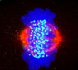
- Immunofluorescent analysis of Phospho-CENP-A pSer7 in a mitotic HeLa cell using a Phospho-CENP-A pSer7 polyclonal antibody (Product # PA5-17195) (green) and a beta-Tubulin monoclonal antibody (red). Phospho-CENP-A signal is localized to bright spots in the metaphase plate. DNA is labeled using a fluorescent blue dye.

- Submitted by
- Invitrogen Antibodies (provider)
- Main image

- Experimental details
- Immunofluorescent analysis of Phospho-CENP-A pSer7 in a mitotic HeLa cell using a Phospho-CENP-A pSer7 polyclonal antibody (Product # PA5-17195) (green) and a beta-Tubulin monoclonal antibody (red). Phospho-CENP-A signal is localized to bright spots in the metaphase plate. DNA is labeled using a fluorescent blue dye.

- Submitted by
- Invitrogen Antibodies (provider)
- Main image

- Experimental details
- Immunofluorescent analysis of Phospho-CENP-A pSer7 in a mitotic HeLa cell using a Phospho-CENP-A pSer7 polyclonal antibody (Product # PA5-17195) (green) and a beta-Tubulin monoclonal antibody (red). Phospho-CENP-A signal is localized to bright spots in the metaphase plate. DNA is labeled using a fluorescent blue dye.
