Explore
 Validate
 Learn
Learn Western blot
 Immunocytochemistry
Antibody data
- Antibody Data
- Antigen structure
- References [16]
- Comments [0]
- Validations
- Western blot [1]
- Immunocytochemistry [1]
- Immunohistochemistry [1]
Submit
Validation data
Reference
Comment
Report error
- Product number
- HPA022040 - Provider product page

- Provider
- Atlas Antibodies
- Proper citation
- Atlas Antibodies Cat#HPA022040, RRID:AB_1846086
- Product name
- Anti-RUNX2
- Antibody type
- Polyclonal
- Description
- Polyclonal Antibody against Human RUNX2, Gene description: runt-related transcription factor 2, Alternative Gene Names: AML3, CBFA1, CCD, CCD1, PEBP2A1, PEBP2aA1, Validated applications: ICC, IHC, WB, Uniprot ID: Q13950, Storage: Store at +4°C for short term storage. Long time storage is recommended at -20°C.
- Reactivity
- Human
- Host
- Rabbit
- Conjugate
- Unconjugated
- Isotype
- IgG
- Vial size
- 100 µl
- Concentration
- 0.1 mg/ml
- Storage
- Store at +4°C for short term storage. Long time storage is recommended at -20°C.
- Handling
- The antibody solution should be gently mixed before use.
Submitted references Biomimetic Intrafibrillar Mineralization of Native Tendon for Soft–Hard Interface Integration by Infiltration of Amorphous Calcium Phosphate Precursors
Digit specific denervation does not inhibit mouse digit tip regeneration
An In Vitro Engineered Osteochondral Model as Tool to Study Osteoarthritis Environment
Breast tumor stiffness instructs bone metastasis via maintenance of mechanical conditioning
Single-cell analysis identifies a key role for Hhip in murine coronal suture development
Atlas of Musculoskeletal Stem Cells with the Soft and Hard Tissue Differentiation Architecture
Chondrocyte-Specific RUNX2 Overexpression Accelerates Post-traumatic Osteoarthritis Progression in Adult Mice
Mandibular dysmorphology due to abnormal embryonic osteogenesis in FGFR2-related craniosynostosis mice
Targeting the gut microbiome to treat the osteoarthritis of obesity
C-type natriuretic peptide analog treatment of craniosynostosis in a Crouzon syndrome mouse model
Time-dependent regulation of morphological changes and cartilage differentiation markers in the mouse pubic symphysis during pregnancy and postpartum recovery
Midface and upper airway dysgenesis in FGFR2-craniosynostosis involves multiple tissue-specific and cell cycle effects
Runx2 contributes to the regenerative potential of the mammary epithelium
BCL11B expression in intramembranous osteogenesis during murine craniofacial suture development
RUNX2 in subtype specific breast cancer and mammary gland differentiation
From shape to cells: mouse models reveal mechanisms altering palate development in Apert syndrome
Chen Y, Zhang Y, Chen X, Huang J, Zhou B, Zhang T, Yin W, Fang C, Yin Z, Pan H, Li X, Shen W, Chen X
Advanced Science 2023;10(34)
Advanced Science 2023;10(34)
Digit specific denervation does not inhibit mouse digit tip regeneration
Dolan C, Imholt F, Yan M, Yang T, Gregory J, Qureshi O, Zimmel K, Sherman K, Smith H, Falck A, Leininger E, Yu L, Brunauer R, Suva L, Gaddy D, Dawson L, Muneoka K
Developmental Biology 2022;486
Developmental Biology 2022;486
An In Vitro Engineered Osteochondral Model as Tool to Study Osteoarthritis Environment
Scalzone A, Cerqueni G, Wang X, Ferreira‐Duarte A, Dalgarno K, Mattioli‐Belmonte M, Gentile P
Advanced Healthcare Materials 2022;12(2)
Advanced Healthcare Materials 2022;12(2)
Breast tumor stiffness instructs bone metastasis via maintenance of mechanical conditioning
Watson A, Grant A, Parker S, Hill S, Whalen M, Chakrabarti J, Harman M, Roman M, Forte B, Gowan C, Castro-Portuguez R, Stolze L, Franck C, Cusanovich D, Zavros Y, Padi M, Romanoski C, Mouneimne G
Cell Reports 2021;35(13):109293
Cell Reports 2021;35(13):109293
Single-cell analysis identifies a key role for Hhip in murine coronal suture development
Holmes G, Gonzalez-Reiche A, Saturne M, Motch Perrine S, Zhou X, Borges A, Shewale B, Richtsmeier J, Zhang B, van Bakel H, Jabs E
Nature Communications 2021;12(1)
Nature Communications 2021;12(1)
Atlas of Musculoskeletal Stem Cells with the Soft and Hard Tissue Differentiation Architecture
Yin Z, Lin J, Yan R, Liu R, Liu M, Zhou B, Zhou W, An C, Chen Y, Hu Y, Fan C, Zhao K, Wu B, Zou X, Zhang J, El‐Hashash A, Chen X, Ouyang H
Advanced Science 2020;7(23)
Advanced Science 2020;7(23)
Chondrocyte-Specific RUNX2 Overexpression Accelerates Post-traumatic Osteoarthritis Progression in Adult Mice
Catheline S, Hoak D, Chang M, Ketz J, Hilton M, Zuscik M, Jonason J
Journal of Bone and Mineral Research 2019;34(9):1676-1689
Journal of Bone and Mineral Research 2019;34(9):1676-1689
Mandibular dysmorphology due to abnormal embryonic osteogenesis in FGFR2-related craniosynostosis mice
Motch Perrine S, Wu M, Stephens N, Kriti D, van Bakel H, Jabs E, Richtsmeier J
Disease Models & Mechanisms 2019;12(5)
Disease Models & Mechanisms 2019;12(5)
Targeting the gut microbiome to treat the osteoarthritis of obesity
Schott E, Farnsworth C, Grier A, Lillis J, Soniwala S, Dadourian G, Bell R, Doolittle M, Villani D, Awad H, Ketz J, Kamal F, Ackert-Bicknell C, Ashton J, Gill S, Mooney R, Zuscik M
JCI Insight 2018;3(8)
JCI Insight 2018;3(8)
C-type natriuretic peptide analog treatment of craniosynostosis in a Crouzon syndrome mouse model
Genetos D, Holmes G, Zhang L, Rivera J, Murphy R, Assouline C, Sullivan L, Oppeneer T, Jabs E
PLOS ONE 2018;13(7):e0201492
PLOS ONE 2018;13(7):e0201492
Time-dependent regulation of morphological changes and cartilage differentiation markers in the mouse pubic symphysis during pregnancy and postpartum recovery
Lammi M, Castelucci B, Consonni S, Rosa V, Sensiate L, Delatti P, Alvares L, Joazeiro P
PLOS ONE 2018;13(4):e0195304
PLOS ONE 2018;13(4):e0195304
Midface and upper airway dysgenesis in FGFR2-craniosynostosis involves multiple tissue-specific and cell cycle effects
Holmes G, O'Rourke C, Perrine S, Lu N, van Bakel H, Richtsmeier J, Jabs E
Development 2018
Development 2018
Runx2 contributes to the regenerative potential of the mammary epithelium
Ferrari N, Riggio A, Mason S, McDonald L, King A, Higgins T, Rosewell I, Neil J, Smalley M, Sansom O, Morris J, Cameron E, Blyth K
Scientific Reports 2015;5(1)
Scientific Reports 2015;5(1)
BCL11B expression in intramembranous osteogenesis during murine craniofacial suture development
Holmes G, van Bakel H, Zhou X, Losic B, Jabs E
Gene Expression Patterns 2015;17(1):16-25
Gene Expression Patterns 2015;17(1):16-25
RUNX2 in subtype specific breast cancer and mammary gland differentiation
McDonald L, Ferrari N, Terry A, Bell M, Mohammed Z, Orange C, Jenkins A, Muller W, Gusterson B, Neil J, Edwards J, Morris J, Cameron E, Blyth K
Disease Models & Mechanisms 2014
Disease Models & Mechanisms 2014
From shape to cells: mouse models reveal mechanisms altering palate development in Apert syndrome
Martínez-Abadías N, Holmes G, Pankratz T, Wang Y, Zhou X, Jabs E, Richtsmeier J
Disease Models & Mechanisms 2013
Disease Models & Mechanisms 2013
No comments: Submit comment
Enhanced validation
- Submitted by
- Atlas Antibodies (provider)
- Enhanced method
- Genetic validation
- Main image

- Experimental details
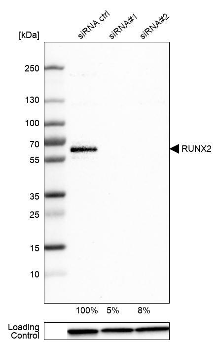
- Western blot analysis in U-251MG cells transfected with control siRNA, target specific siRNA probe #1 and #2, using Anti-RUNX2 antibody. Remaining relative intensity is presented. Loading control: Anti-GAPDH.
- Sample type
- Human
- Protocol
- Protocol
Supportive validation
- Submitted by
- Atlas Antibodies (provider)
- Main image
- Experimental details
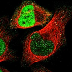
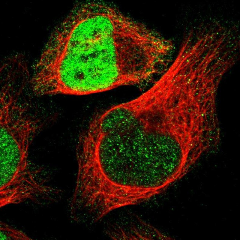
- Immunofluorescent staining of human cell line U-2 OS shows localization to nucleoplasm.
- Sample type
- Human
Supportive validation
- Submitted by
- Atlas Antibodies (provider)
- Enhanced method
- Orthogonal validation
- Main image
- Experimental details
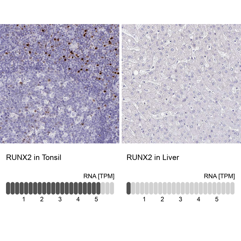
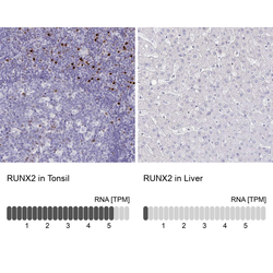
- Immunohistochemistry analysis in human tonsil and liver tissues using HPA022040 antibody. Corresponding RUNX2 RNA-seq data are presented for the same tissues.
- Sample type
- Human
- Protocol
- Protocol