Explore
 Validate
 Learn
Learn Western blot
 ELISA
ELISAAntibody data
- Antibody Data
- Antigen structure
- References [2]
- Comments [0]
- Validations
- Western blot [1]
- Immunohistochemistry [2]
- Flow cytometry [1]
Submit
Validation data
Reference
Comment
Report error
- Product number
- ABIN453492 - Provider product page

- Provider
- antibodies-online
- Product name
- anti-Abl-Interactor 1 (ABI1) (N-Term), (AA 87-117) antibody
- Antibody type
- Polyclonal
- Antigen
- KLH conjugated synthetic peptide between 87~117 amino acids from the N-terminal region of Human ABI1
- Description
- Affinity chromatography on Protein A
- Reactivity
- Human
- Host
- Rabbit
- Epitope
- N-Term,AA 87-117
- Vial size
- 0.4 mL
- Concentration
- 0.25 mg/mL
- Storage
- Store the antibody undiluted at 2-8°C for one month or (in aliquots) at -20°C for longer.
- Handling
- Avoid repeated freezing and thawing.
Submitted references Rac interacts with Abi-1 and WAVE2 to promote an Arp2/3-dependent actin recruitment during chlamydial invasion.
Abelson interactor protein-1 positively regulates breast cancer cell proliferation, migration, and invasion.
Carabeo RA, Dooley CA, Grieshaber SS, Hackstadt T
Cellular microbiology 2007 Sep;9(9):2278-88
Cellular microbiology 2007 Sep;9(9):2278-88
Abelson interactor protein-1 positively regulates breast cancer cell proliferation, migration, and invasion.
Wang C, Navab R, Iakovlev V, Leng Y, Zhang J, Tsao MS, Siminovitch K, McCready DR, Done SJ
Molecular cancer research : MCR 2007 Oct;5(10):1031-9
Molecular cancer research : MCR 2007 Oct;5(10):1031-9
No comments: Submit comment
Supportive validation
- Submitted by
- antibodies-online (provider)
- Main image
- Experimental details
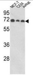
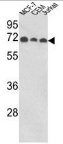
- Western blot analysis of ABI1 Antibody (N-term) (AP17861PU-N) in MCF-7, CEM, Jurkat cell line lysates (35ug/lane). ABI1 (arrow) was detected using the purified Pab.
Supportive validation
- Submitted by
- antibodies-online (provider)
- Main image
- Experimental details
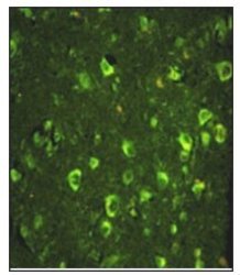
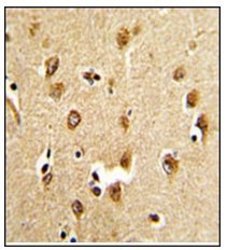
- Immunofluorescence analysis of ABI1 Antibody (N-term) with paraffin-embedded human brain tissue. 0.05 mg/ml primary antibody was followed by FITC-conjugated goat anti-rabbit lgG (whole molecule). FITC emits green fluorescence.

- Submitted by
- antibodies-online (provider)
- Main image
- Experimental details
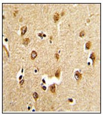
- Immunohistochemistry analysis in Formalin Fixed, Paraffin Embedded Human brain tissue stained with ABI1 Antibody (N-term) Cat.-No AP17861PU-N which was peroxidase-conjugated to the secondary antibody, followed by DAB staining. This data demonstrates the use of this antibody for immunohistochemistry; clinical relevance has not been evaluated.
Supportive validation
- Submitted by
- antibodies-online (provider)
- Main image
- Experimental details
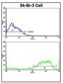
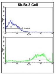
- Flow cytometric analysis of SK-Br-3 cells using ABI1 Antibody (N-term)(bottom histogram) compared to a negative control cell (top histogram). FITC-conjugated goat-anti-rabbit secondary antibodies were used for the analysis.