Explore
 Validate
 Learn
Learn Western blot
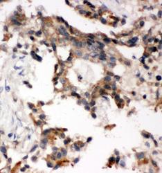
 Immunohistochemistry
Antibody data
- Antibody Data
- Antigen structure
- References [0]
- Comments [0]
- Validations
- Immunohistochemistry [1]
Submit
Validation data
Reference
Comment
Report error
- Product number
- PA1001 - Provider product page

- Provider
- Boster Biological Technology
- Product name
- Anti-SSH3BP1/ABI1 Antibody
- Antibody type
- Polyclonal
- Description
- Polyclonal antibody for SSH3BP1/ABI1 detection. Host: Rabbit.Size: 100μg/vial. Tested applications: WB, IHC-P, IF. Reactive species: Human. SSH3BP1/ABI1 information: Molecular Weight: 55081 MW; Subcellular Localization: Cytoplasm . Nucleus . Cell projection, lamellipodium . Cell projection, filopodium . Cell projection, growth cone . Cell junction, synapse, postsynaptic cell membrane, postsynaptic density . Cytoplasm, cytoskeleton . Localized to protruding lamellipodia and filopodia tips. Also localized to neuronal growth cones and synaptosomes. May shuttle from the postsynaptic densities to the nucleus (By similarity); Tissue Specificity: Widely expressed, with highest expression in brain.
- Reactivity
- Human, Mouse, Rat
- Host
- Rabbit
- Vial size
- 100μg/vial
- Concentration
- Add 0.2ml of distilled water will yield a concentration of 500ug/ml.
- Storage
- At -20°C for one year. After reconstitution, at 4°C for one month. It can also be aliquoted and stored frozen at -20°C for a longer time. Avoid repeated freezing and thawing.
- Handling
- Add 0.2ml of distilled water will yield a concentration of 500ug/ml.
No comments: Submit comment
Supportive validation
- Submitted by
- Boster Biological Technology (provider)
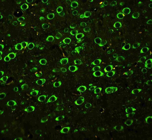
- Main image

- Experimental details
- IF analysis of ABI using anti- ABI antibody (PA1001) ABI was detected in paraffin-embedded section of rat brain tissues. Heat mediated antigen retrieval was performed in citrate buffer (pH6, epitope retrieval solution ) for 20 mins. The tissue section was blocked with 10% goat serum. The tissue section was then incubated with 1μg/mL rabbit anti- ABI Antibody (PA1001) overnight at 4°C. DyLight®488 Conjugated Goat Anti-Rabbit IgG (BA1127) was used as secondary antibody at 1:100 dilution and incubated for 30 minutes at 37°C. Visualize using a fluorescence microscope and filter sets appropriate for the label used.
- Additional image