Explore
 Validate
 Learn
Learn Western blot
 Flow cytometry
Antibody data
- Antibody Data
- Antigen structure
- References [3]
- Comments [0]
- Validations
- Flow cytometry [1]
- Other assay [1]
Submit
Validation data
Reference
Comment
Report error
- Product number
- MAB229 - Provider product page

- Provider
- Novus Biologicals
- Product name
- Mouse Monoclonal CD30/TNFRSF8 Antibody
- Antibody type
- Monoclonal
- Description
- Protein A or G purified from hybridoma culture supernatant. Detects human CD30/TNFRSF8 in direct ELISAs and Western blots. In direct ELISAs, less than 1% cross-reactivity with recombinant mouse CD30, recombinant human (rh) CD27, and rhCD40 is observed.
- Reactivity
- Human
- Host
- Mouse
- Conjugate
- Unconjugated
- Isotype
- IgG
- Vial size
- 500 ug
- Storage
- Use a manual defrost freezer and avoid repeated freeze-thaw cycles. 12 months from date of receipt, -20 to -70 degreesC as supplied. 1 month, 2 to 8 degreesC under sterile conditions after reconstitution. 6 months, -20 to -70 degreesC under sterile conditions after reconstitution.
Submitted references Molecular Signatures of Dengue Virus-Specific IL-10/IFN-γ Co-producing CD4 T Cells and Their Association with Dengue Disease.
Cytoplasmic Pin1 expression is increased in human cutaneous melanoma and predicts poor prognosis.
Selective redox regulation of cytokine receptor signaling by extracellular thioredoxin-1.
Tian Y, Seumois G, De-Oliveira-Pinto LM, Mateus J, Herrera-de la Mata S, Kim C, Hinz D, Goonawardhana NDS, de Silva AD, Premawansa S, Premawansa G, Wijewickrama A, Balmaseda A, Grifoni A, Vijayanand P, Harris E, Peters B, Sette A, Weiskopf D
Cell reports 2019 Dec 24;29(13):4482-4495.e4
Cell reports 2019 Dec 24;29(13):4482-4495.e4
Cytoplasmic Pin1 expression is increased in human cutaneous melanoma and predicts poor prognosis.
Chen X, Liu X, Deng B, Martinka M, Zhou Y, Lan X, Cheng Y
Scientific reports 2018 Nov 15;8(1):16867
Scientific reports 2018 Nov 15;8(1):16867
Selective redox regulation of cytokine receptor signaling by extracellular thioredoxin-1.
Schwertassek U, Balmer Y, Gutscher M, Weingarten L, Preuss M, Engelhard J, Winkler M, Dick TP
The EMBO journal 2007 Jul 11;26(13):3086-97
The EMBO journal 2007 Jul 11;26(13):3086-97
No comments: Submit comment
Supportive validation
- Submitted by
- Novus Biologicals (provider)
- Main image
- Experimental details
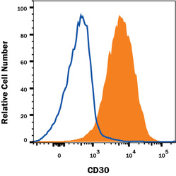
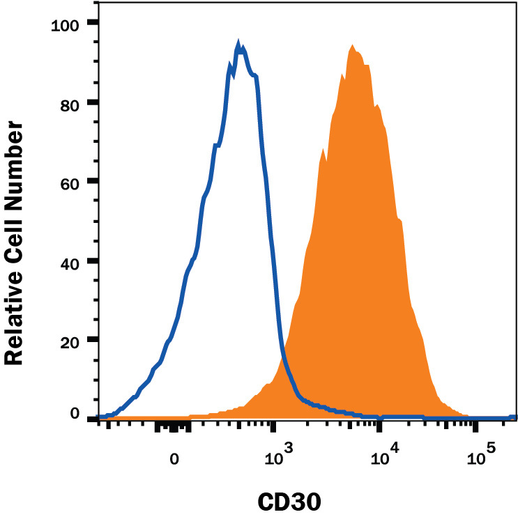
- Detection of CD30/TNFRSF8 in Jurkat Human Cell Line by Flow Cytometry. Jurkat human acute T cell leukemia cell line was stained with Mouse Anti-Human CD30/TNFRSF8 Monoclonal Antibody (Catalog # MAB229, filled histogram) or isotype control antibody (Catalog # MAB0041, open histogram), followed by Phycoerythrin-conjugated Anti-Mouse IgG Secondary Antibody (Catalog # F0102B). View our protocol for Staining Membrane-associated Proteins.
Supportive validation
- Submitted by
- Novus Biologicals (provider)
- Main image
- Experimental details
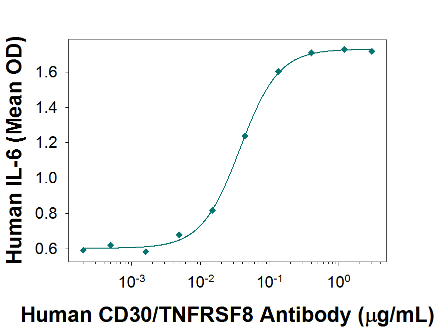
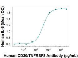
- Human CD30/TNFRSF8 Antibody Enhances IL-6 Secretion in HDLM-2 Cells. Human CD30/TNFRSF8 Antibody Enhances IL-6 Secretion in HDLM-2 Cells.