 Explore
 Validate
 Learn
Learn Western blot
 ELISA
ELISAAntibody data
- Antibody Data
- Antigen structure
- References [0]
- Comments [0]
- Validations
- Western blot [3]
Submit
Validation data
Reference
Comment
Report error
- Product number
- LS-C18809 - Provider product page

- Provider
- LSBio
- Proper citation
- LifeSpan Cat#LS-C18809, RRID:AB_2244045
- Product name
- CCND1 / Cyclin D1 Antibody LS-C18809
- Antibody type
- Polyclonal
- Description
- Delipidated and defibrinated
- Reactivity
- Human
- Host
- Rabbit
- Storage
- Store at 4°C or -20°C. Avoid freeze-thaw cycles.
No comments: Submit comment
Supportive validation
- Submitted by
- LSBio (provider)
- Enhanced method
- Genetic validation
- Main image

- Experimental details
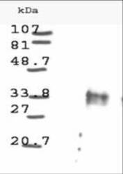
- Western blot analysis is shown using Anti-Cyclin D1 antibody to detect Human Cyclin D1 present in asynchronous HN30 cell lysates. HN30 cells, are from head and neck cancer cells that over express cyclin B1 and D1. Comparison to a molecular weight marker indicates a band of ~34 kD corresponding to the expected molecular weight for the protein (arrowhead). The blot was incubated with a 1:500 dilution of the antibody at room temperature. Detection occurred using a 1:10000 of HRP conjugated Go.

- Submitted by
- LSBio (provider)
- Enhanced method
- Genetic validation
- Main image

- Experimental details
- Western blot analysis is shown using Anti-Cyclin D1 antibody to detect Human Cyclin D1 present in asynchronous HN30 cell lysates. HN30 cells, are from head and neck cancer cells that over express cyclin B1 and D1. Comparison to a molecular weight marker indicates a band of ~34 kD corresponding to the expected molecular weight for the protein (arrowhead). The blot was incubated with a 1:500 dilution of the antibody at room temperature. Detection occurred using a 1:10000 of HRP conjugated Go.

- Submitted by
- LSBio (provider)
- Enhanced method
- Genetic validation
- Main image
- Experimental details
- Western blot analysis is shown using Anti-Cyclin D1 antibody to detect Human Cyclin D1 present in asynchronous HN30 cell lysates. HN30 cells, are from head and neck cancer cells that over express cyclin B1 and D1. Comparison to a molecular weight marker indicates a band of ~34 kD corresponding to the expected molecular weight for the protein (arrowhead). The blot was incubated with a 1:500 dilution of the antibody at room temperature. Detection occurred using a 1:10000 of HRP conjugated Go.
